MICROSCOPE SLIDE or SEM STUB PREPARATION for OCTOCORAL SCLERITES or OTHER INVERTEBRATE SPICULES
Compiled by Gary Williams and Courtney Mattison
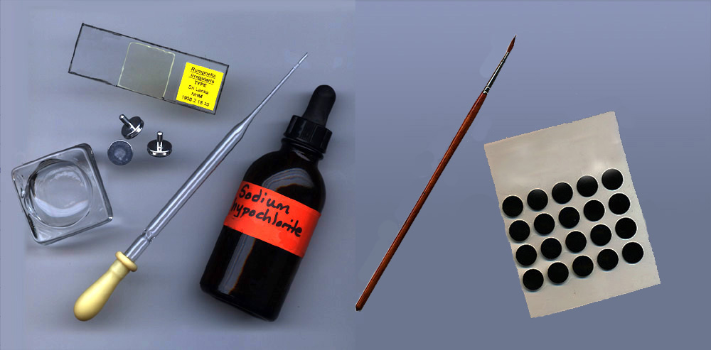
1. Put a small amount of tissue (about 5 cubic millimeters or less) in a circular watch glass (ca. 30-35 mm diameter).
2. Add 2 drops of fresh household bleach (sodium hypochlorite - NaOCl) to the tissue, using an eyedropper or pipette.
3. Macerate for a few minutes until a sufficient quantity of sclerites have been dissociated from the tissue sample. Remove pieces of excess tissue.
4. Fill the watch glass with clean water. Allow all of the sclerites to sink to the bottom, then pipette off the liquid. Repeat this washing process one or two more times.
5. Fill the watch glass with 95% ethanol. Allow all the sclerites to sink to the bottom, then carefully pipette off the fluid. Fill the watch glass with 95% ethanol once again. A clockwise circular flow in the ethanol generated by gentle puffs from the pipette on the inside edge of the watch glass will result in sclerites densely concentrated in the center of the watch glass.
6. Allow the sclerites to sink, then pipette a quantity of sclerites in ethanol from the watch glass to the center of a glass microscope slide. Air-dry completely until the sclerites look like dry powder on the microscope slide.
7. FOR MICROSCOPE SLIDES:
* NOTE: for temporary slides, use GLYCEROL instead of a permanent mounting medium such as CYTOSEAL.
8. FOR SEM STUBS: