|
||||

|
ENTOMOLOGY
RESOURCES
|
ARACHNIDA |
EMBIIDINA |
FAUNAL PROJECTS |
FOSSIL INSECTS |
HYMENOPTERA |
MECOPTERA |
NEUROPTERA |
Atlas of Entelegynae
| To search the atlas for any keyword press: ctrl + f Figure 1. Right spinnerets of Hypochilus pococki female from Haywood Co., North Carolina , USA . A. Spinnerets and cribellum, overview. B. ALS. C. PMS. D. PLS. AC = aciniform gland spigot(s), MAP = major ampullate gland spigot(s), MS = PLS modified spigot (s), PI = piriform gland spigots. Scale bars: A = 500µm, B–D = 100µm. Figure 2. Details of female spinnerets. A–D. Hypochilus pococki from Haywood Co., North Carolina , USA . A. Cribellum and left ALS. Arrow to intermediate ALS segment. B. Cribellar spigots, close up. C. Right ALS, MAP field: two MAP spigots larger than the rest. D. Right PLS: arrows to two (possibly three, one broken shaft) apical spigots larger than the AC. E. Ectatosticta sp. from Shaanxi Province, China , right PLS: arrows to two apical spigots smaller than the AC. AC = aciniform gland spigot(s), MAP = major ampullate gland spigot(s), PI = piriform gland spigots. Scale bars: A = 400µm, B = 10µm, C, D = 50µm, D = 201µm, E = 100µm. Figure 3. Spinnerets of female Filistata insidiatrix from Siena , Italy . A. Overview, right. B. Right ALS. C. PMS. D. Right PLS. E. Right PMS. F. Right PLS, detail of PC. AC = aciniform gland spigot(s), MAP = major ampullate gland spigot(s), mAP = minor ampullate gland spigot(s), PC = paracribellar spigot(s). Scale bars: A = 300μm, B–E = 50μm, F = 20μm. Figure 4. Left male spinnerets of Filistatainsidiatrix from Barcelona , Spain . A. Spinneret overview. B. ALS. C. PMS. D. PLS. AC = aciniform gland spigot(s), MAP = major ampullate gland spigot(s), mAP = minor ampullate gland spigot(s), PC = paracribellar spigot(s), PI = piriform gland spigot(s). Scale bars: A = 200µm, B–D = 50µm. Figure 5. Details of spinnerets of Filistata insidiatrix female from Siena , Italy . A. Cribellum. B–C. Cribellar spigots. D. Left ALS. Note modified setae posteriorly. E. Left ALS, detail of modified setae. F. Right ALS showing three MAP, one of them among the PI. Note absence of tartipores. MAP = major ampullate gland spigot(s), PI = piriform gland spigot(s). Scale bars: A, D = 100μm, B = 5μm, C = 3μm, E = 30μm, F = 40μm. Figure 6. Left spinnerets of Kukulcaniahibernalis female from Alachua Co., Florida , USA . A. Overview. B. ALS. C. PMS, anterior view, with mAP hidden. D. PLS. Note absence of tartipores. AC = aciniform gland spigot(s), MAP = major ampullate gland spigot(s), PC = paracribellar spigot(s), PI = piriform gland spigot(s). Scale bars: A, D = 200µm, B, D = 100µm. Figure 7. Right spinnerets of female Hickmania troglodytes from Mole Creek Cave , Tasmania , Australia . A. Spinnerets, overview. B. ALS. C. PMS. D. PLS. Arrows to CY spigots. AC = aciniform gland spigot(s), CY = cylindrical gland spigot(s), MAP = major ampullate gland spigot(s), mAP = minor ampullate gland spigot(s), MS = PLS modified spigot, PC = paracribellar spigot(s), TP = tartipore(s). Scale bars: A = 1.0mm, B = 300μm, C = 100μm, D = 500μm. Figure 8. Right spinnerets of male Hickmania troglodytes from Mole Creek Cave , Tasmania , Australia . A. Spinnerets, overview. B. ALS. C. PMS: Question marks to presumed AC and mAP spigots with larger shafts. D. PLS. AC = aciniform gland spigot(s), MAP = major ampullate gland spigot(s), mAP = minor ampullate gland spigot(s), MS = PLS modified spigot, PC = paracribellar spigot(s), PI = piriform gland spigot(s), TP = tartipore(s). Scale bars: A = 1.0mm, B, D = 300μm, C = 100μm. Figure 9. Right male and female PLS of Hickmania troglodytes from Mole Creek Cave , Tasmania , Australia . A–B. Female PLS showing CY spigots forming an external-anterior row, and two sizes of AC spigots. C–D. Male PLS showing two sizes of AC spigots. AC = aciniform gland spigot(s), CY = cylindrical gland spigot(s), MS = PLS modified spigot, TP = tartipore(s). Scale bars: A, C = 100μm, B = 50μm, D = 40μm. Figure 10. Details of spinnerets of female Hickmania troglodytes from Mole Creek Cave , Tasmania , Australia . A–B. Cribellum. C–F. Right spinnerets. C. ALS: the marginal PI are larger. D. ALS showing marginal pair of MAP and tartipore. E. PMS showing mAP with accompanying tartipore. F. PMS showing long paracribellars, and two size classes of AC. AC = aciniform gland spigot(s), CY = cylindrical gland spigot(s), MAP = major ampullate gland spigot(s), mAP = minor ampullate gland spigot(s), PC = paracribellar spigot(s), PI = piriform gland spigot(s), TP = tartipore(s). Scale bars: A = 500μm, B = 10μm, C, D = 100μm, E, F = 50μm. Figure 11. Female spinnerets of Thaida peculiaris from Puerto Blest , Argentina . A. Spinnerets and cribellum, overview. B–D. Right spinnerets. B. ALS. C. PMS. D. PLS. AC = aciniform gland spigot(s), CY (and arrows) = cylindrical gland spigot(s), MAP = major ampullate gland spigot(s), mAP = minor ampullate gland spigot(s), PC = paracribellar spigot(s), MS = PLS modified spigot, PI = piriform gland spigot(s), TP = tartipore(s). Scale bars: A = 1.0mm, B–D = 100μm. Figure 12. Right male spinnerets of Thaida peculiaris from Cautin , Chile . A. Spinnerets and cribellum, overview. B. ALS. C. PMS. D. PLS. AC = aciniform gland spigot(s), MAP = major ampullate gland spigot(s), mAP = minor ampullate gland spigot(s), MS = PLS modified spigot nubbin, N = nubbins, PC = paracribellar spigot and nubbins, TP = tartipore(s). Parenthetical notations, i.e., (PC) and (MS) refer to nubbins. Scale bars: A = 500μm, B = 100μm, C, D = 50μm. Figure 13. Details of female spinnerets of Thaida peculiaris from Puerto Blest , Argentina . A. Cribellum. B. Cribellar spigots. C. Right ALS, MAP and TP. D. Right PMS, posterior view. E–F. Apex of right PLS: asterisk to intermediate paracribellar shaft with only partial sculpturing. AC = aciniform gland spigot(s), CY and arrows = cylindrical gland spigot(s), MAP = major ampullate gland spigot(s), mAP = minor ampullate gland spigot(s), MS = PLS modified spigot, N = nubbin, PC = paracribellar spigot(s), TP = tartipore(s). Scale bars: A = 500μm, B = 10μm, C, E, F = 50μm, D = 100μm. Figure 14. Spinning organs of female filistatids and austrochilids. A, B. Thaida peculiaris from Osorno , Chile . A. Left ALS, MAP and tartipore. B. PLS apex. C, D. Kukulcaniahibernalis from Alachua Co., Florida , USA . C. PMS, showing PC spigots. D. Cribellum. E. Filistatinella sp. from Arroyo Seco, California, USA, clavate cribellar spigots. MS = PLS modified spigot, PC = paracribellar spigot(s), TP = tartipores. Scale bars: A–C = 20µm, D = 100µm, E = 3µm. Figure 15. Right spinnerets of Gradungula sorenseni female from Saltwater Forest , New Zealand . A. Spinnerets, overview. B. ALS. C. PMS. D. PLS. AC = aciniform gland spigot(s), MAP = major ampullate gland spigot(s), PI = piriform gland spigot(s), TP = tartipore(s). Scale bars: A = 500μm, B, C = 200μm, D = 300μm. Figure 16. Right spinnerets of Gradungula sorenseni male from Saltwater Forest , New Zealand . A. Spinnerets, overview. B. ALS. C. PMS. D. PLS. AC = aciniform gland spigot(s), MAP = major ampullate gland spigot(s), PI = piriform gland spigot(s), TP = tartipore(s). Scale bars: A = 500μm, B, D = 100μm, C = 50μm. Figure 17. Details of right spinnerets of Gradungula sorenseni female from Saltwater Forest , New Zealand . A. ALS, PI. B–C. ALS, MAP field. D–E. PMS. F. PLS, large AC at top, small in center. AC = aciniform gland spigot(s), MAP = major ampullate gland spigot(s), PI = piriform gland spigot(s), TP = tartipore(s). * denotes small orifices. Scale bars: A, C = 30μm, B, F = 50μm, D = 40μm, E = 10μm. Figure 18. Female spinnerets of Ariadna boesenbergi from Buenos Aires , Argentina . A. Spinnerets, anterior-ventral view. B. Spinnerets, posterior-ventral view. C, D. Right ALS. E. Left PMS. F. Left PLS. AC = aciniform gland spigot(s), MAP = major ampullate gland spigot(s), mAP = minor ampullate gland spigot(s), PI = piriform gland spigot(s). Scale bars: A = 200 μm, B = 50 μm, C, D = 200 μm, E, F = 20 μm. Figure 19. Male spinnerets of Ariadna boesenbergi from Buenos Aires , Argentina . A. Spinnerets. B. Right ALS. C. Right PMS. D. Right PLS (arrow to reduced AC spigot, present in the left PLS). AC = aciniform gland spigot(s), MAP = major ampullate gland spigot(s), mAP = minor ampullate gland spigot(s), PI = piriform gland spigot(s). Scale bars: A = 200 μm, B = 40 μm, C = 20 μm, D = 20 μm. Figure 20. Spinnerets of Archaeidae and Araneoidea. A–D. Archaea workmani immature from Ranomafana , Madagascar . A. Spinnerets. B. Left ALS: the tartipore is probably hidden in the fold around MAP. C. PMS. D. Left PLS. E. Araneus diadematus female from Ontario , Canada , left ALS (arrow to fold separating MAP field). F. Crassanapis chilensis, female from Osorno , Chile , right ALS (inverted, arrows to fold separating MAP field). AC = aciniform gland spigot(s), MAP = major ampullate gland spigot(s), mAP = minor ampullate gland spigot(s), N = nubbin, PI = piriform gland spigot(s), TP = tartipore(s). Scale bars: A = 50μm, B–D = 10μm, E = 100μm, F = 20μm. Figure 21. Right spinnerets of female Archaea workmani from Ranomafana , Madagascar . A. Spinneret overview. B. ALS. C. PMS. D. PLS. AC = aciniform gland spigot(s), CY = cylindrical gland spigot(s), MAP = major ampullate gland spigot(s), mAP = minor ampullate gland spigot(s), N = nubbin, PI = piriform gland spigot(s), TP = tartipore(s). Scale bars: A = 100μm, B = 40μm, C, D = 30μm. Figure 22. Spinnerets of male Archaea workmani from Ranomafana , Madagascar , left, inverted. A. Overview. B. ALS. C. PMS. D. PLS. AC = aciniform gland spigot(s), MAP = major ampullate gland spigot(s), mAP = minor ampullate gland spigot(s), N = nubbin of posterior MAP. Scale bars: A = 100μm, B = 40μm, C, D = 20μm. Figure 22. Spinnerets of male Archaea workmani from Ranomafana , Madagascar , left, inverted. A. Overview. B. ALS. C. PMS. D. PLS. AC = aciniform gland spigot(s), MAP = major ampullate gland spigot(s), mAP = minor ampullate gland spigot(s), N = nubbin of posterior MAP. Scale bars: A = 100μm, B = 40μm, C, D = 20μm. Figure 23. Left spinnerets of Huttonia wiltoni female from Orongorongo , New Zealand . A. Overview. B. ALS. C. PMS. D. PLS. AC = aciniform gland spigot(s), CY and arrows = cylindrical gland spigot(s), MAP = major ampullate gland spigot(s), N = nubbin, PI = piriform gland spigot(s), TP = tartipore(s). Scale bars: A = 300μm, B, C = 50μm, D = 100μm. Figure 24. Left spinnerets of Huttonia wiltoni male from Orongorongo , New Zealand . A. Overview. B. ALS. C. PMS. D. PLS. AC = aciniform gland spigot(s), MAP = major ampullate gland spigot(s), N = nubbin, PI = piriform gland spigot(s), TP = tartipore(s). Scale bars: A = 200μm, B = 40μm, C, D = 50μm. Figure 25. Spinnerets of female Mimetus hesperus from Tampico , Mexico . A. Left overview. B. Left ALS. Note very short PI bases. C. Right PMS. D. Left PLS. AC = aciniform gland spigot(s), CY = cylindrical gland spigot(s), MAP = major ampullate gland spigot(s), mAP = minor ampullate gland spigot(s), N = nubbin (in white, of posterior ALS MAP, in black, of PMS mAP), PI = piriform gland spigot(s), TP = tartipore (s). Scale bars: A = 115µm, B = 30µm, C, D = 25µm. Figure 26. Left spinnerets of male Mimetus hesperus from Baboquivari Mts. , Arizona , USA . A. Overview. B. ALS. C. PMS. D. PLS. AC = aciniform gland spigot(s), MAP = major ampullate gland spigot(s), mAP = minor ampullate gland spigot(s), N = nubbin (in white, of posterior ALS MAP, in black, of PMS mAP), PI = piriform gland spigot(s). Scale bars: A = 100µm, B, C = 30µm, D = 25µm. Figure 27. Spinnerets of female Oecobiusnavus from Richmond , California , USA . A. Overview. Note divided cribellum, elongate PLS and enlarged anal tubercle. B. Left ALS. C. Left PMS, with median field of AC. D. Left PLS, with numerous AC. AC = aciniform gland spigot(s), CY = cylindrical gland spigot(s), MAP = major ampullate gland spigot(s), mAP = minor ampullate gland spigot(s), PI = piriform gland spigot(s), TP = tartipore(s). Scale bars: A = 200µm, B = 20µm, C = 15µm, D = 75µm. Figure 28. Spinnerets of male Oecobiusnavus from Washington D.C, USA . A. Overview of spinnerets. B. Left ALS. C. Left PMS, anterior to bottom. D. Right PLS, with only AC spigots. AC = aciniform gland spigot(s), MAP = major ampullate gland spigot(s), mAP = minor ampullate gland spigot(s), PI = piriform gland spigot(s), TP = tartipore(s). Scale bars: A = 200µm, B,C = 20µm, D = 100µm. Figure 29. Spinnerets of female Uroctea sp. from Garies , South Africa . A. Left ALS. B. Left PMS, with no apparent mAP. C. Base of left PLS, with numerous AC. D. Left PLS. AC = aciniform gland spigot(s), CY = cylindrical gland spigot(s), MAP = major ampullate gland spigot(s), PI = piriform gland spigot(s), TP = tartipore(s). Scale bars: A, B = 75µm, C = 150µm, D = 430µm. Figure 30. Spinnerets of male Uroctea sp. from Garies , South Africa . A. Spinneret overview. Note elongate PLS and enlarged anal tubercle. B. Left ALS, showing mostly MAP spigots. C. Left PMS, probably with only AC spigots. D. Left PLS showing only AC spigots. AC = aciniform gland spigot(s), MAP = major ampullate gland spigot(s), PI = piriform gland spigot(s). Scale bars: A = 430µm, B, C = 60µm, D = 200µm. Figure 31. Spinnerets of female Eresus cf. cinnaberinus from Igrherm , Morocco . A. Overview. B. Left ALS: larger spigots (arrows) are presumably MAP, smaller PI. C. Right PMS: larger spigots are presumably mAP and CY, smaller AC. D. Left PLS (inverted). AC = aciniform gland spigot(s) , MAP = major ampullate gland spigot(s), mAP = minor ampullate gland spigot(s), MS = PLS modified spigot PI = piriform gland spigot(s). Scale bars: A = 1.0mm, B–D = 200μm. Figure 32. Right spinnerets of male Eresus cf. cinnaberinus from Mistras , Greece . A. Spinneret overview. Arrow to intermediate ALS segment. B. ALS, arrows to MAP. C. PMS, arrows to mAP. D. PLS, arrows to vestigial spigots accompanying the MS . AC = aciniform gland spigot(s), MAP = major ampullate gland spigot(s), mAP = minor ampullate gland spigot(s), MS = PLS modified spigot, N = nubbin(s), PI = piriform gland spigot(s). Scale bars: A = 300μm, B, C = 100μm, D = 50μm. Figure 33. Spinnerets of Eresidae. A–F. Eresus cf. cinnaberinus. A–D. Female from Igrherm , Morocco . E, F. Male from Mistras , Greece . A. Cribellar spigots. B. Detail of right female ALS, showing MAP (arrows) interspersed among PI spigots; the large tartipores correspond to MAP, the small to PI. C. Detail of right female PMS: the ampullate shafts are characteristically sculptured. D. Detail of female base of right PLS (inverted), showing MS and accompanying spigots (arrows). E. Detail of right male PMS: one of the mAP is much larger than the rest. F. Detail of base of right male PLS (inverted), showing MS and vestigial accompanying spigots (arrows). G–J. Stegodyphus mimosarum female from Phinda , South Africa . G. Cribellar spigots. H. Detail of right ALS showing MAP (arrows) interspersed among PI spigots; the large tartipores correspond to MAP, the small to PI. I. Apical detail of right PLS: several nubbins among the AC. J. Detail of base of right PLS: showing MS and two accompanying spigots. AC = aciniform gland spigot(s), MAP = major ampullate gland spigot(s), mAP = minor ampullate gland spigot(s), MS = PLS modified spigot , N = nubbin(s), PI = piriform gland spigot(s). Scale bars: A, G = 5 μ m, B, D = 100 μ m, C, H = 50 μ m, E, F, I = 40 μ m, J = 30 μ m. Figure 34. Spinnerets of female Eresidae. A, C. Eresus sandaliatus from Silkeborg, Denmark. B, E. Eresus cinnaberinus from Fiesch Wallis , Switzerland . D, F. Stegodyphusmimosaurum, from Fort Hill , Malawi . A. Cribellum. B. Left ALS, detail. C. Strobilate cribellar spigots. D. Left PMS, detail. Note characteristic sculpturing of ampullate shaft. E. PMS. F. Right PLS apex. AC = aciniform gland spigots, MAP = major ampullate gland spigot(s), mAP = minor ampullate gland spigot(s), PI = piriform gland spigot(s), TP = tartipore(s). Scale bars: A = 300µm, B = 25µm, C = 4.3µm, D = 15µm, E = 60µm, F = 20µm. Figure 35. Spinnerets of female Stegodyphusmimosarum from Fort Hill , Malawi . A. Left spinneret overview. B. Right ALS. Smaller spigots are presumably PI. C. Left PMS. D. Right PLS. AC = aciniform gland spigots, CY = cylindrical gland spigot(s), MAP = major ampullate gland spigot(s), mAP = minor ampullate gland spigot(s), N = nubbin(s), PI = piriform gland spigot(s), TP = tartipore(s). Scale bars: A = 200µm, B = 60µm, C, D = 43µm. Figure 36. Right spinnerets of male Stegodyphusmimosarum from Fort Hill , Malawi . A. Overview (inset enlarged in D). B. ALS. C. PMS. D. PLS, inset to MS (area marked in A). AC = aciniform gland spigots, MAP = major ampullate gland spigot(s) , MS = PLS modified spigot, PI = piriform gland spigot(s), TP = tartipore(s). Scale bars: A = 150µm, B = 43µm, C, D = 30µm. Figure 37. Right spinnerets of female Stegodyphus mimosarum from Phinda , South Africa . A. Overview of cribellum and spinnerets. B. ALS. Arrows to MAP. C. PMS. D. PLS. CY = cylindrical gland spigot, MAP = major ampullate gland spigot(s), mAP = minor ampullate gland spigot(s), MS = PLS modified spigot, PI = piriform gland spigot(s), TP = tartiportes. Scale bars: A = 400μm, B, D = 100μm, C = 50μm. Figure 38. Spinnerets of Nicodamidae and Araneidae. A, B, D. Megadictynathilenii. A, D. Female from Hick's Bay , New Zealand . B. Juvenile from Cherry Bay , New Zealand . C, E. Female Argiopeargentata from Baja California, Mexico. A. Apex of left PLS showing PC, MS and AC spigots. B. PMS, showing PC and AC spigots. C. Triplet of AG and FL spigots at apex of PLS. D. Cribellum. E. Left PLS. AC = aciniform gland spigot(s), AG = aggregate gland spigots, CY = cylindrical gland spigot(s), FL = flagelliform gland spigot, MS = PLS modified spigot, PC = paracribellar spigot(s). Scale bars: A = 30µm, B = 15µm, C = 43μm, D, E = 150µm. Figure 39. Left spinnerets of female Megadictynathilenii from Hicks Bay , New Zealand . A. Overview. B. ALS. C. PMS. Note posterior mAP. PC spigots are hidden. D. PLS. AC = aciniform gland spigots, MAP = major ampullate gland spigot(s), mAP = minor ampullate gland spigot(s), MS = PLS modified spigot, PC = paracribellar spigot(s), PI = piriform gland spigot(s), TP = tartipore(s). Scale bars: A = 300µm, B = 75µm, C = 100µm, D = 150µm. Figure 40. Left spinnerets of male Megadictynathilenii from Orongorongo , New Zealand . A. Overview. B. ALS. Note difference in size between anterior and posterior MAP. C. PMS. Note posterior mAP and anterior encircling line of PC spigot nubbins. D. PLS. Note apical nubbins of MS and PC. AC = aciniform gland spigot(s), MAP = major ampullate gland spigot(s), mAP = minor ampullate gland spigot(s), MS = PLS modified spigot nubbin, PC = paracribellar spigot nubbins, PI = piriform gland spigot(s). Scale bars: A = 254µm, B = 75µm, C, D = 100µm. Figure 41. Right spinnerets of female Nicodamus mainae from Coalseam Park , Western Australia , Australia . A. Spinneret overview. B. ALS. C. PMS. D. PLS. AC = aciniform gland spigot(s), CY = cylindrical gland spigot(s), MAP = major ampullate gland spigot(s), mAP = minor ampullate gland spigot(s), PI = piriform gland spigot(s), TP = tartipore(s). Scale bars: A = 500μm, B–D = 100μm. Figure 42. Left spinnerets (inverted) of male Nicodamus mainae from Bush Bay , Western Australia , Australia . A. Spinneret overview. B. ALS. C. PMS, nubbin posterior to mAP is absent on right spinneret. D. PLS, apical nubbin is absent on right spinneret. AC = aciniform gland spigot(s), MAP = major ampullate gland spigot(s), mAP = minor ampullate gland spigot(s), N = nubbins, PI = piriform gland spigot(s), TP = tartipore(s). Scale bars: A = 500μm, B = 50μm, C, D = 100μm. Figure 43. Spinnerets of female Menneuscamelus from Zululand , South Africa . A. Cribellum and PMS. B. Left ALS. C. Left PMS. D. Left PLS. AC = aciniform gland spigot(s), CY = cylindrical gland spigot(s), MAP = major ampullate gland spigot(s), mAP = minor ampullate gland spigot(s), MS = PLS modified spigot, PC = paracribellar spigot(s), PI = piriform gland spigot(s), TP = tartipore(s). Scale bars: A = 500μm, B = 100μm, C, D = 200μm. Figure 44. Spinneret details of female Deinopoidea. A, B. Octonobaoctonaria from St. Charles Co., Missouri , USA . A. Spinnerets, anteroventral view. B. PLS. C–E. Menneuscamelus from Zululand , South Africa . C. ALS MAP. D. PLS apex, showing modified spigot. E. PLS detail. AC = aciniform gland spigot(s), CY = cylindrical gland spigot(s), MAP = major ampullate gland spigot(s), MS = PLS modified spigot, TP = tartipore(s). Scale bars: A = 500μm, B = 50μm, C–E = 20μm. Figure 45. Spinnerets of female Orbiculariae. A–C. Uloborustrilineatus from Pipeline Road, Panama . A. PLS. B. ALS. C. PMS. D. Deinopisspinosus from Alachua Co., Florida , USA , PMS paracribellars. AC = aciniform gland spigot(s), CY = cylindrical gland spigot(s), MAP = major ampullate gland spigot(s), mAP = minor ampullate gland spigot(s), MS = PLS modified spigot, N = nubbin(s), PC = paracribellar spigot(s), PI = piriform gland spigot(s), TP = tartipore(s). Scale bars: A = 100μm, B–D = 50μm. Figure 46. Spigots of female Phyxelididae. A. Phyxelidatanganensis from Amani , Tanzania , apex of left PLS showing CY, MS and AC spigots. B, C. Namaquarachnetropata from Grootvadersbosch , South Africa . B. PLS apex. C. PMS anterior, with PC labelling ends of encircling row of PC spigots with flattened bases. D. Vytfutiapallens from Sarawak , Malaysia , left PMS apex. AC = aciniform gland spigot(s), CF = cuticular finger, CY = cylindrical gland spigot(s), mAP = minor ampullate gland spigot(s), MS = PLS modified spigot, PC = paracribellar spigot(s). Scale bars: A, B = 20µm, C = 40µm, D = 15µm. Figure 47. Spinnerets of female Vytfutiapallens from Sarawak , Malaysia . A. Spinneret overview. B. Left ALS. Note nubbin of MAP spigot and cuticular fold mesad of MAP spigot. C. Right PMS with PC labeling ends of encircling row of PC spigots. Note flattened bases of PC spigots. D. Right PLS. AC = aciniform gland spigot(s), CY = cylindrical gland spigot(s), MAP = major ampullate gland spigot(s), mAP = minor ampullate gland spigot(s), MS = PLS modified spigot, N = nubbin(s), PC = paracribellar spigot(s), PI = piriform gland spigot(s). Scale bars: A = 150µm, B = 35µm, C = 25µm, D = 20µm. Figure 48. Spinnerets of male Vytfutiapallens from Sarawak , Malaysia . A. Right spinneret overview. B. Right ALS showing nubbin of posterior MAP spigot. C. Left PMS, showing nubbins of PC spigots. D. Left PLS showing nubbins of MS and PC spigots. AC = aciniform gland spigot(s), MAP = major ampullate gland spigot(s), mAP = minor ampullate gland spigot(s), N = nubbins, PC = paracribellar spigot nubbins on PMS, PI = piriform gland spigot(s). Scale bars: A = 100µm, B, D = 30µm, C = 25µm. Figure 49. Left spinnerets of female Phyxelidatanganensis from Amani , Tanzania . A. Overview. B. ALS. C. PMS. Lines point to ends of encircling row of PC spigots. D. PLS. Arrow to stout seta near PLS apex. AC = aciniform gland spigot(s), CY = cylindrical gland spigot(s), MAP = major ampullate gland spigot(s), mAP = minor ampullate gland spigot(s), MS = PLS modified spigot, N = nubbin, PC = paracribellar spigot(s), PI = piriform gland spigot(s), TP = tartipore. Scale bars: A = 150µm, B, D = 43µm, C = 46µm. Figure 50. Right spinnerets of male Phyxelidatanganensis from Amani , Tanzania . A. Overview. B. ALS. C. PMS. Note large TP. D. PLS, showing nubbin of MS at apex . AC = aciniform gland spigot(s), MAP = major ampullate gland spigot(s), mAP N = minor ampullate gland spigot nubbin, MS = nubbin of PLS modified spigot, PC = nubbins of PC spigots, PI = piriform gland spigot(s), TP = tartipore(s). Scale bars: A = 150µm, B = 30µm, C, D = 20µm. Figure 51. Spinnerets of female Titanoeca americana from Johnson Co., Missouri , USA . A. Right spinneret overview. B. Left ALS. MAP are sunken into broken cuticle. C. Left PMS. D. Right PLS. AC = aciniform gland spigot(s), CY = cylindrical gland spigot(s), MAP = major ampullate gland spigot(s), mAP = minor ampullate gland spigot(s), PC = paracribellar spigot(s), PI = piriform gland spigot(s). Scale bars: A = 200µm, B = 30µm, C = 20µm, D = 43µm. Figure 52. Titanoeca spinnerets. A. Female Titanoecanigrella from Cave Creek , Arizona , USA , PLS apex. B–D. Male Titanoeca americana from Johnson Co., Missouri , USA , left. B. ALS. C. PMS showing AC spigots and large nubbin of mAP. D. PLS, showing AC spigots and nubbins of apical and basal PC. AC = aciniform gland spigot(s), CY = cylindrical gland spigot(s), N = nubbin(s), PC = paracribellar spigot(s). Scale bars: A = 43µm, B, D = 30µm, C = 25µm. Figure 53. Spinnerets of female Goeldia sp. from Zapallar , Chile . A. Left overview. B. Right ALS. C. Right PMS. Note absence of PC. D. Left PLS. Basal and apical PC labeled. AC = aciniform gland spigot(s), CY = cylindrical gland spigot(s), MAP = major ampullate gland spigot(s), mAP = minor ampullate gland spigot(s), PC = paracribellar spigot(s), PI = piriform gland spigot(s), TP = tartipore. Scale bars: A = 150µm, B, D = 30µm, C = 20µm. Figure 54. Spinnerets of male Goeldia sp. from Zapallar , Chile . A. Right ALS. B. Right PLS. Note nubbin of apical PC. C, D. PMS. Nubbin is probably vestige of mAP. C. Lateral. D. Posterior. AC = aciniform gland spigot(s), MAP = major ampullate gland spigot(s), N = nubbin(s), PC = nubbin of paracribellar spigot, PI = piriform gland spigot(s), TP = tartipore(s). Scale bars: A–D = 25µm. Figure 55. Spinnerets of female Titanoecidae. A, C. Titanoecanigrella from Cave Creek, Arizona , USA . A. Right ALS. C. Left PLS. B, D. Titanoeca americana from Johnson Co., Missouri , USA . B. Right PLS apex, showing PC spigot. D. Right PLS. E, F. Goeldia spp. E. Zapallar , Chile , left PLS. F. Valparaiso , Chile , right PLS. AC = aciniform gland spigot(s), CY = cylindrical gland spigot(s), MAP = major ampullate gland spigot(s), PC = paracribellar spigot(s), PI = piriform gland spigot(s). Scale bars: A, B = 20µm, C = 43µm, D = 60µm, E = 43µm, F = 20µm. Figure 56. Spinneret details. A–C. Aebutina binotata female from Tarapuy , Ecuador . Black line to MS spigots and black arrows to flanking spigots of triad. A. Overview. B. Left PLS. C. Left PLS, base of apical segment showing presumed flanking spigots of MS triad. D. Psechrus sp. male from Tham Lot Cave , Thailand , left PLS. AC = aciniform gland spigot(s), MS = PLS modified spigot, N = nubbin of MS spigot. Scale bars: A = 250 μ m, B = 100 μ m, C = 15 μ m, D = 60µm. Figure 57. Spinnerets of female Aebutina binotata from Rio Cuyabeno , Ecuador . A. Overview. B. Right ALS. C. Right PMS. D. Left PLS: note basal MS and flanking spigots of triad (arrows). AC = aciniform gland spigot(s), CY = cylindrical gland spigot(s), MAP = major ampullate gland spigot(s), mAP = minor ampullate gland spigot(s), MS = PLS modified spigot(s), PI = piriform gland spigot(s), TP = tartipores . Scale bars: A = 200µm, B = 30µm, C = 25µm, D = 43µm. Figure 58. Spinnerets of male Aebutina binotata from Tarapuy , Ecuador . A. Right overview. B. Right ALS. C. PMS. D. Left PLS. Note anterior basal nubbins of MS and flanking spigots of triad. AC = aciniform gland spigot(s), MAP = major ampullate gland spigot and nubbin, mAP = minor ampullate gland spigot(s), mAP N = minor ampullate gland spigot nubbins, MS = nubbin of PLS modified spigot, N = nubbins of anterobasal PLS spigots . Scale bars: A = 75µm, B = 20µm, C = 43µm, D = 30µm. Figure 59. Spinnerets of female Dictynaarundinacea from Lingby , Denmark . A. Overview. B. Left ALS. C. Left PMS, anterior to left. Note PC spigots encircling top and left side of image, and multiple PC spigots emerging from single large base at bottom. D. Left PLS. AC = aciniform gland spigot(s), CY = cylindrical gland spigot(s), MAP = major ampullate gland spigot(s), mAP = minor ampullate gland spigot(s), MS = PLS modified spigot, N = nubbin(s), PC = paracribellar spigot(s), PI = piriform gland spigot(s), TP = tartipore. Scale bars: A = 200µm, B–D = 20µm. Figure 60. Spinnerets of male Dictynaarundinacea from Lingby , Denmark . A. Overview. B. Left ALS. C. Left PMS, showing PC nubbins encircling spinneret anterolaterally. D. Left PLS, showing tartipores amid AC spigot field. AC = aciniform gland spigot(s), MAP = major ampullate gland spigot(s), mAP = minor ampullate gland spigot(s), N = nubbin(s), N(MS) = nubbin of PLS modified spigot, PC = paracribellar spigot nubbins, PI = piriform gland spigot(s), TP = tartipore(s), TP (MAP) = tartipore of MAP from previous instar. Scale bars: A = 200µm, B, D = 20µm, C = 50µm. Figure 61. Left spinnerets of female Nigmalinsdalei from San Francisco , California , USA . A. Overview. B. ALS. C. PMS. Note PC spigots encircling spinneret, several that have multiple shafts emerging from a common base. D. PLS. AC = aciniform gland spigot(s), CY = cylindrical gland spigot(s), MAP = major ampullate gland spigot(s), mAP = minor ampullate gland spigot(s), MS = PLS modified spigot, N = nubbin of MAP, PC = paracribellar spigot(s), PI = piriform gland spigot(s), TP = tartipore(s). Scale bars: A = 59µm, B = 15µm, C, D = 20µm. Figure 62. Left spinnerets of male Nigmalinsdalei from San Francisco , California , USA . A. Overview. B. ALS. C. PMS. D. PLS. AC = aciniform gland spigot(s), MAP = major ampullate gland spigot(s), mAP = minor ampullate gland spigot(s), N = nubbins of ALS MAP and PLS PC and MS, PC = nubbins of PMS paracribellar spigots, PI = piriform gland spigot(s), TP = tartipore. Scale bars: A = 60µm, B–D = 15µm. Figure 63. Left spinnerets of female Lathyshumilis from Kent , United Kingdom . A. Overview. B. ALS. C. PMS. D. PLS. AC = aciniform gland spigot(s), CY = cylindrical gland spigot(s), MAP = major ampullate gland spigot(s), mAP = minor ampullate gland spigot(s), MS = PLS modified spigot, N = nubbin, PC = paracribellar spigot(s), PI = piriform gland spigot(s), TP = tartipore. Scale bars: A = 60µm, B = 15µm, C, D = 10µm. Figure 64. Right spinnerets of male Lathyshumilis from Kent , United Kingdom . A. Overview. B. ALS. C. PMS. D. PLS. AC = aciniform gland spigot(s), MAP = major ampullate gland spigot(s), mAP = minor ampullate gland spigot(s), MS = PLS modified spigot nubbin, N = nubbin of posterior MAP, PC = paracribellar spigot nubbins, PI = piriform gland spigot(s), TP = tartipore. Scale bars: A = 43µm, B = 15µm, C, D = 10µm. Figure 65. Right spinnerets of female Tricholathys sp. from San Francisco , California , USA . A. Overview. B. ALS. C. PMS apex. D. PLS. AC = aciniform gland spigot(s), CY = cylindrical gland spigot(s), MAP = major ampullate gland spigot(s), mAP = minor ampullate gland spigot(s), N = nubbin(s), PC = paracribellar spigot(s), PI = piriform gland spigot(s), TP = tartipore(s). Scale bars: A = 75µm, B, D = 15µm, C = 10µm. Figure 66. Spinning organs of Dictynidae. A, F, G. Tricholathysspiralis from Lenore Lake, Washington, USA. A. Left female PMS PC with multiple shafts emerging from wide base. F. Right female PMS. G. Left male PMS. B. Nigmalinsdalei female from San Francisco , California , USA , left PMS showing PC with multiple shafts per base encircling spinneret. C. Dictynaarundinacea female from Lingby , Denmark , PLS apex. D. Lathyshumilis female from Kent , United Kingdom , cribellum. E. Nigmalinsdalei, female from San Francisco , California , USA , cribellar spigots. AC = aciniform gland spigot(s), CY = cylindrical gland spigot(s), mAP = minor ampullate gland spigot(s), MS = PLS modified spigot, N = nubbin of paracribellar spigot(s), PC = paracribellar spigot(s). Scale bars: A = 7.5µm, B = 15µm, C = 10µm, D = 43µm, E = 6µm, F = 25µm, G = 20µm. Figure 67. Left spinnerets of female Neolanadalmasi from Waipoua Forest , New Zealand . A. Overview. B. ALS. C. PMS. D. PLS. AC = aciniform gland spigot(s), CY = cylindrical gland spigot(s), MAP = major ampullate gland spigot(s), mAP = minor ampullate gland spigot(s), MS = PLS modified spigot, PC = paracribellar spigot(s), PI = piriform gland spigot(s), TP = tartipore(s). Scale bars: A = 200µm, B = 43µm, C = 25µm, D = 60µm. Figure 68. Spinneret details of female Neolanidae and Stiphidiidae. A. Neolanadalmasi from Waipoua Forest , New Zealand , PLS apex. B, D. Stiphidionfacetum from Bundeena , New South Wales , Australia . B. PMS. D. PLS apex. C. Pillaragriswoldi from Barrington Tops, New South Wales , Australia , PLS apex. AC = aciniform gland spigot(s), mAP = minor ampullate gland spigot(s), MS = PLS modified spigot, PC = paracribellar spigot(s). Scale bars: A = 100µm, B–D = 20µm. Figure 69. Spinnerets of female Stiphidionfacetum from Bundeena , New South Wales , Australia . A. Overview. B. Left ALS. C. Left PMS. D. Left PLS. CY = cylindrical gland spigot(s), MAP = major ampullate gland spigot(s), mAP = minor ampullate gland spigot(s), MS = PLS modified spigot(s), PC = paracribellar spigot(s), PI = piriform gland spigot(s). Scale bars: A = 200µm, B, C = 50µm, D = 100µm. Figure 70. Spinnerets of male Stiphidionfacetum from Whalipu , New Zealand . A. Overview. B. Right ALS. C. PMS. D. Right PLS. AC = aciniform gland spigot(s), mAP = minor ampullate gland spigot(s), N = nubbins of PC and MS on PLS apex, PC = paracribellar spigot nubbins on PMS, TP = tartipore(s). Scale bars: A = 200µm, B = 30µm, C, D = 60µm. Figure 71. Spinnerets of female Pillara griswoldi from Barrington Tops, New South Wales , Australia . A. Overview. B. Left ALS. C. Left PMS. D. Left PLS. AC = aciniform gland spigot(s), CY = cylindrical gland spigot(s), MAP = major ampullate gland spigot(s), mAP = minor ampullate gland spigot(s), MS = PLS modified spigot, PC = paracribellar spigot(s), PI = piriform gland spigot(s). Scale bars: A = 200µm, B, C = 20µm, D = 50µm. Figure 72. Spinnerets of Pillara griswoldi from Barrington Tops, New South Wales , Australia . A. Strobilate cribellar spigots of female. B. Male right ALS. C. Male right PMS. D. Male right PLS. AC = aciniform gland spigot(s), MAP = major ampullate gland spigot(s), mAP = minor ampullate gland spigot(s), N = nubbins of PC and MS on PLS apex, PC = paracribellar spigot nubbins on PMS, PI = piriform gland spigot(s). Scale bars: A = 10µm, B = 50µm, C = 20µm, D = 100µm. Figure 73. Left spinnerets of female Neoramia sana from Dunedin , New Zealand . A. Overview. B. ALS. C. PMS. D. PLS. AC = aciniform gland spigot(s), CY = cylindrical gland spigot(s), MAP = major ampullate gland spigot(s), mAP = minor ampullate gland spigot(s), MS = PLS modified spigot, PC = paracribellar spigot(s), PI = piriform gland spigot(s), TP = tartipore(s). Scale bars: A = 200µm, B, D = 60µm, C = 30µm. Figure 74. Left spinnerets of male Neoramia sana from Dunedin , New Zealand . A. Overview. B. ALS. C. PMS. D. PLS. MAP = major ampullate gland spigot(s), mAP = minor ampullate gland spigot(s), N = nubbins, PC = PMS paracribellar spigot nubbins. Scale bars: A = 200µm, B, D = 60µm, C = 30µm. Figure 75. Spinnerets of female Manihongaitahu from Kaituna Valley , New Zealand . A. Left overview. B. Left ALS. C. Right PMS, showing median row of PC spigots. D. Left PLS. AC = aciniform gland spigot(s), CY = cylindrical gland spigot(s), MAP = major ampullate gland spigot(s), mAP = minor ampullate gland spigot(s), MS = PLS modified spigot, PC = paracribellar spigot(s), PI = piriform gland spigot(s). Scale bars: A = 150µm, B, C = 43µm, D = 60µm. Figure 76. Spinnerets of male Manihongaitahu from Kaituna Valley , New Zealand . A. Left overview. B. Right ALS. C. Left PMS. D. Left PLS. AC = aciniform gland spigot(s), mAP = minor ampullate gland spigot(s), N = nubbins (of PC spigots on PMS, of PC and MS spigots on PLS). Scale bars: A = 150µm, B = 43µm, C = 30µm, D = 60µm. Figure 77. Right spinnerets of female Metaltellasimoni from Riverside , California , USA . A. Overview. B. ALS. C. PMS, showing median field of PC spigots. D. PLS. AC = aciniform gland spigot(s), CY = cylindrical gland spigot(s), MAP = major ampullate gland spigot(s), mAP = minor ampullate gland spigot(s), MS = PLS modified spigot, PC = paracribellar spigot(s), PI = piriform gland spigot(s), TP = tartipore(s). Scale bars: A = 200µm, B, D = 60µm, C = 43µm. Figure 78. Right spinnerets of male Metaltellasimoni from Riverside , California , USA . A. Overview. B. ALS. C. PMS. Note median field of PC nubbins. D. PLS. AC = aciniform gland spigot(s), MAP = major ampullate gland spigot(s), mAP = minor ampullate gland spigot(s), N = nubbins of MS and PLS PC, PC = nubbins of PMS PC, TP = tartipore(s). Scale bars: A = 150µm, B = 43µm, C = 30µm, D = 60µm. Figure 79. Female spinnerets of Desis formidabilis from Cape Peninsula , South Africa . A. Spinneret overview. B. Left ALS. C. Left PMS. D. Left PLS. AC = aciniform gland spigot(s), CY = cylindrical gland spigot(s), MAP = major ampullate gland spigot(s), mAP = minor ampullate gland spigot(s), MS = PLS modified spigot, PI = piriform gland spigot(s), TP = tartipore(s). Scale bars: A = 1.0mm, B = 100µm, C, D = 200µm. Figure 80. Male spinnerets of Desis formidabilis from Cape Peninsula , South Africa . A. Spinnerets (arrow to tracheal spiracle). B. Left ALS, arrow to modified setae. C. Left PMS. D. Left PLS. AC = aciniform gland spigot(s), MAP = major ampullate gland spigot(s), mAP = minor ampullate gland spigot(s), MS = PLS modified spigot, PI = piriform gland spigot(s), TP = tartipore(s). Scale bars: A = 1.0mm, B 100µm, C = 300µm, D = 200µm. Figure 81. Spinnerets and spigots of Desis formidabilis from Cape Peninsula , South Africa . A. Female right ALS, note the dense setal cover. B. Detail of female right ALS spinning field, arrows to modified setae. C. Detail of female left ALS, the small mound (N?) between MAP and TP may correspond with the nubbin of a MAP. D. Same, male left ALS. E. Detail of male left PMS. F. Detail of female left PLS. AC = aciniform gland spigot(s), CY = cylindrical gland spigot(s), MAP = major ampullate gland spigot(s), mAP = minor ampullate gland spigot(s), MS = PLS modified spigot, N = nubbin, PI = piriform gland spigot(s), TP = tartipore(s). Scale bars: A = 300µm, B—E = 50µm, F = 100µm. Figure 82. Spinnerets. A, B, F. Neoramia sana from Dunedin , New Zealand . A. Apex of female PLS. B. Nubbin on male PMS with PC spigot. F. Cribellum. C, G. Metaltellasimoni female from Riverside , California , USA . C. Apex of PLS. G. Cribellum. D, E. Manihongaitahu female from Kaituna Valley , New Zealand . D. Right PMS apex. E. Left PLS apex. AC = aciniform gland spigot(s), CF = cuticular finger, mAP = minor ampullate gland spigot(s), MS = PLS modified spigot, PC = paracribellar spigot(s). Scale bars: A = 20µm, B = 10µm, C = 6µm, D, E = 15µm, F = 150µm, G = 250µm. Figure 83. Left spinnerets of female Matachiaaustralis from Dunedin , New Zealand . A. Overview. B. ALS. C. Left and right PMS. Note numerous PC spigots with one shaft per base. D. PLS. AC = aciniform gland spigot(s), MAP = major ampullate gland spigot(s), mAP = minor ampullate gland spigot(s), MS = PLS modified spigot, PC = paracribellar spigot(s), PI = piriform gland spigot(s). Scale bars: A = 150µm, B = 30µm, C = 60µm, D = 43µm. Figure 84. Female spinnerets of Phryganoporus candidus from Canberra , Australia . A. Spinnerets. B. Left ALS. C. Left PMS. D. Left PLS. AC spigots with shafts labelled, of others only bases remain. Note common base of PC and MS. AC = aciniform gland spigot(s), CY = cylindrical gland spigot(s), MAP = major ampullate gland spigot(s), mAP = minor ampullate gland spigot(s), MS = PLS modified spigot, PC = paracribellar spigot(s), PI = piriform gland spigot(s). Scale bars: A = 200µm, B, D = 50µm, C = 20µm. Figure 85. Male spinnerets of Phryganoporus candidus from Canberra , Australia . A. Spinnerets. B. Right ALS. C. Right PMS. Note PC nubbins. D. Right PLS. Note nubbins of PC and of common base to PC and MS. AC = aciniform gland spigot(s), MAP = major ampullate gland spigot(s), mAP = minor ampullate gland spigot(s), N = nubbins of posterior MAP on ALS, PC and MS on PLS, PC = paracribellar spigot nubbins on PMS, TP = tartipores. Scale bars: A = 200µm, B–D = 50µm Figure 86. Left female spinnerets of Badumnalonginqua from Maui , Hawaii , USA . A. Overview. B. ALS. C. PMS. D. PLS. AC = aciniform gland spigot(s), CY = cylindrical gland spigot(s), MAP = major ampullate gland spigot(s), mAP = minor ampullate gland spigot(s), MS = PLS modified spigot, PC = paracribellar spigot(s), PI = piriform gland spigot(s) , TP = tartipore(s). Scale bars: A = 200µm, B, D = 100µm, C = 50µm. Figure 87. Female spinnerets of Desidae. A, E. Matachiaaustralis from Dunedin , New Zealand . B. Phryganoporus candidus from Canberra, Australia. C, D, F. Badumnalonginqua from Maui , Hawaii , USA . A. Left PLS apex, showing PC and MS amid AC spigots. B. PLS apex, showing PC and MS with common base, and AC with sharp-edged base. C. PMS showing mid-spinning-field PC with multiple shafts from each base. D. Left PLS apex, showing PC and MS with common base, and AC with sharp-edged base. E, F. Cribella. AC = aciniform gland spigot(s), mAP = minor ampullate gland spigot(s), MS = PLS modified spigot, PC = paracribellar spigot(s). Scale bars: A = 15µm, B–D = 20µm, E, F = 200µm. Figure 88. Left spinnerets of female Amaurobiusfenestralis from Tisvilde , Denmark . A. Overview. B. ALS. C. PMS. D. PLS, showing PC and MS arising from common base. AC = aciniform gland spigot(s), CY = cylindrical gland spigot(s), MAP = major ampullate gland spigot(s), mAP = minor ampullate gland spigot(s), MS = PLS modified spigot, PC = paracribellar spigot(s), PI = piriform gland spigot(s), TP = tartipore(s). Scale bars: A = 200µm, B, D = 50µm, C = 20µm. Figure 89. Spinnerets of female Callobiusbennetti from Hampshire Co., West Virginia , USA . A. Right overview. B. Right ALS, showing wide margin around spinning field. C. PMS. D. Right PLS. AC = aciniform gland spigot(s), CY = cylindrical gland spigot(s), MAP = major ampullate gland spigot(s), mAP = minor ampullate gland spigot(s), MS = PLS modified spigot, PC = paracribellar spigot(s), PI = piriform gland spigot(s), TP = tartipore(s). Scale bars: A = 200µm, B–D = 100µm. Figure 90. Spinnerets of female Pimus sp. from Mendocino Co., California , USA . A. Left overview. B. Left ALS. C. Left PMS. Debris obscures the spinning field, but comparison with the male suggests that there are two mAP and one PC. D. Right PLS. AC = aciniform gland spigot(s), CY = cylindrical gland spigot(s), MAP = major ampullate gland spigot(s), mAP = minor ampullate gland spigot(s), MS = PLS modified spigot , PC = paracribellar spigot(s), PI = piriform gland spigot(s). Scale bars: A = 75µm, B = 25µm, C = 43µm, D = 30µm. Figure 91. Spinnerets of male Pimus sp. from Mendocino Co., California , USA . A. Left overview. B. Right ALS, showing MAP, nubbin and tartipore. C. Left PMS. Note two large spigots that may be mAP. D. Left PLS showing nubbins of two PC and one MS spigots. AC = aciniform gland spigot(s), MAP = major ampullate gland spigot(s), mAP = minor ampullate gland spigot(s), MS = nubbin of PLS modified spigot, N = MAP nubbin, PC = paracribellar spigot nubbins, PI = piriform gland spigot(s), TP = tartipore(s). Scale bars: A = 150µm, B, C = 20µm, D = 30µm. Figure 92. Spinnerets of female Retiro sp. from Lima , Peru . A. Left overview. B. Right ALS, with only one MAP and nubbin. The second nubbin may be a PI with broken shaft. C. Right PMS. D. Right PLS. Apical MS spigot shaft broken off. AC = aciniform gland spigot(s), CY = cylindrical gland spigot(s), MAP = major ampullate gland spigot(s), mAP = minor ampullate gland spigot(s), MS = PLS modified spigot , N = nubbin(s), PI = piriform gland spigot(s), TP = tartipore(s). Scale bars: A = 150µm, B = 30µm, C = 43µm, D = 60µm. Figure 93. Left spinnerets of male Retiro sp. from Lima , Peru . A. Overview. B. ALS. C. Left and right PMS. D. PLS, showing AC spigots and MS nubbin. AC = aciniform gland spigot(s), MAP = major ampullate gland spigot(s), mAP = minor ampullate gland spigot(s), MS = PLS modified spigot nubbin , N = nubbin of posterior MAP, PI = piriform gland spigot(s). Scale bars: A = 150µm, B, C = 43µm, D = 60µm. Figure 94. Female spinnerets of Macrobunus multidentatus from Chiloé , Chile . A. Spinnerets. B. Colulus. C. Left ALS. D. Left PMS. E. Left PLS. AC = aciniform gland spigot(s), CY = cylindrical gland spigot(s), MAP = major ampullate gland spigot(s), mAP = minor ampullate gland spigot(s), TP = tartipore. Scale bars: A = 200 μ m, B–E = 50 μ m. Figure 95. Male spinnerets of Macrobunus multidentatus from Chiloé , Chile . A. Spinnerets. B. Right ALS. C. Right PMS. D. Right PLS. AC = aciniform gland spigot(s), MAP = major ampullate gland spigot(s), mAP = minor ampullate gland spigot(s), N = nubbin(s), TP = tartipore. Scale bars: A = 200 μ m, B, C, D = 50 μ m. Figure 96. Female spinnerets. A, B, E. Retiro sp. from Lima, Peru. A. Cribellum. B. Close up of strobilate cribellar spigots. E. Apex of right PLS. C. Callobiusbennetti from Hampshire Co., West Virginia , USA , PMS apex, showing PC bases with single and multiple shafts. D. Pimus sp. from Mendocino Co., California , USA , apex of right PLS. F–H. Pimus napa from Napa Co., California , USA , right spinnerets. F. ALS. G. PMS. H. PLS. AC = aciniform gland spigot(s), CY = cylindrical gland spigot(s), MAP = major ampullate gland spigot(s), mAP = minor ampullate gland spigot(s), MS = PLS modified spigot, PC = paracribellar spigot(s), TP = tartipore . Scale bars: A = 150µm, B = 6µm, C, D = 10µm, E = 15µm, F, H = 50µm, G = 40µm. Figure 97. Female cribellar spinning organs. A, G. Acanthoctenus sp. from Changuinola , Panama . B, E. Psechrus sp. from Tham Lot Cave , Thailand . C, F. Uduba sp. from Ranomafana , Madagascar . D. Raecius jocquei from Appouesso , Côte d'Ivoire . A–D. Cribella. E—G. Details of cribellar spigots. Scale bars: A = 150µm, B = 430µm, C = 200µm, D = 201µm, E, G = 7.5µm, F = 25µm. Figure 98. Spinnerets of female Tengella radiata from Guanacaste , Costa Rica . A. Right spinnerets, overview. B. Left ALS. C. Left PMS: Arrows to larger ‘AC’. D. Right PLS. Arrows to larger ‘AC’. Inset: MS and two flanking spigots (arrows). AC = aciniform gland spigot(s), CY = cylindrical gland spigot(s), MAP = major ampullate gland spigot(s), mAP = minor ampullate gland spigot(s), MS = PLS modified spigot, TP = tartipore(s). Scale bars: A = 100μm, B, D = 200μm, C = 100μm. Figure 99. Left spinnerets of male Tengella radiata from Limón , Costa Rica . A. Spinnerets, overview. B. ALS. C. PMS. D. PLS. E. Detail of PMS, anterior view. F. Detail of PLS: arrows to larger ‘AC’. Note the intermediate spigot-seta to the right of the MS nubbin. AC = aciniform gland spigot(s), MAP = major ampullate gland spigot(s), mAP = minor ampullate gland spigot(s), MS = nubbin of PLS modified spigot, N = MAP nubbin, TP = tartipore(s). Scale bars: A = 500μm, B–E = 100μm, F = 50μm. Figure 100. Spinnerets of female Tengellidae and Zoropsidae. A–C. Tengellaradiata from La Selva , Costa Rica . A. Left PLS apex. B. Right PLS apex. C. Right ALS, MAP with broken shafts and PI. D—G. Zoropsisspinimana from Barcelona , Spain . D. Left PLS apex. E. PMS apex. F. Left PMS, posterior view. G. Left PLS, ectal. AC = aciniform gland spigot(s), CY = cylindrical gland spigot(s), MAP = major ampullate gland spigot(s), mAP = minor ampullate gland spigot(s), MS = PLS modified spigot, PI = piriform gland spigot(s), TP = tartipore(s). Scale bars: A, B, D, F = 100μm, C, E = 50μm, G = 20μm. Figure 101. Spinnerets of female Zorocrates cf. mistus from Los Llanos, Chiapas , Mexico . A. Cribellum. B. Cribellar spigots. C. Detail of left PLS, showing MS and nubbins of the two accompanying spigots (arrows). D. Detail of right PLS, showing MS and the two accompanying spigots (arrows), one of them arising from the MS base. AC = aciniform gland spigot(s), MS = PLS modified spigot. Scale bars: A = 50µm, B = 10µm, C, D = 50µm. Figure 102. Spinnerets of female Zorocrates cf. mistus from Los Llanos, Chiapas , Mexico . A. Spinnerets. B. Right ALS. C. Left PMS, posterior view. D. Left PMS, detail. E. Right PLS. F. Left PLS (arrow to scar similar to a nubbin). AC = aciniform gland spigot(s), CY = cylindrical gland spigot(s), MAP = major ampullate gland spigot(s), mAP = minor ampullate gland spigot(s), MS = PLS modified spigot, TP = tartipore(s). Scale bars: A = 1.0mm, B, D—F = 100µm, C = 200µm. Figure 103. Spinnerets of male Zorocrates sp. from Big Bend National Park , Texas , USA . A. Spinnerets. B. Left ALS (MAP shaft broken). C. Right PMS, posterior view. D. Right PMS. E. Left PLS. F. Left PLS, detail of nubbins from MS and its accompanying spigots. G. Vestiges of cribellum. AC = aciniform gland spigot(s), MAP = major ampullate gland spigot(s), mAP = minor ampullate gland spigot(s), N = nubbin(s), TP = tartipore(s). Scale bars: A = 500µm, B, D—F = 100µm, C = 150µm, G = 10µm. Figure 104. Right spinnerets of female Uduba sp. from Ranomafana , Madagascar . A. Overview. B. ALS. C. PMS. D. PLS. Inset: MS and two flanking spigots. AC = aciniform gland spigot(s), CY = cylindrical gland spigot(s), MAP = major ampullate gland spigot(s), mAP = minor ampullate gland spigot(s), MS = PLS modified spigot, PI = piriform gland spigot(s), TP = tartipore(s). Scale bars: A = 440µm, B, D = 100µm, C = 150µm. Figure 105. Right spinnerets of female Raecius jocquei paratype. A. Overview. B. ALS. C. PMS. D. PLS. AC = aciniform gland spigot(s), CY = cylindrical gland spigot(s), MAP = major ampullate gland spigot(s), mAP = minor ampullate gland spigot(s), MS = PLS modified spigot, PI = piriform gland spigot(s), TP = tartipore(s). Scale bars: A = 150 μ m, B–D = 43 μ m. Figure 106. Left spinnerets of male Raecius jocquei paratype. A. Overview. B. ALS. C. PMS. D. PLS. AC = aciniform gland spigot(s), MAP = major ampullate gland spigot(s), mAP = minor ampullate gland spigot(s), mAP N = minor ampullate gland nubbin, MS N = nubbin of PLS modified spigot, N = MAP nubbin, PI = piriform gland spigot(s), TP = tartipore(s). Scale bars: A = 150 μ m, B–D = 43 μ m. Figure 107. Spinnerets of female Poaka graminicola from Lincoln , New Zealand . A. Left overview. B. Right ALS. C. PMS. D. Right PLS. AC = aciniform gland spigot(s), CY = cylindrical gland spigot(s), MAP = major ampullate gland spigot(s) and nubbin, mAP = minor ampullate gland spigot(s), MS = PLS modified spigot, PC = paracribellar spigot, PI = piriform gland spigot(s). Scale bars: A = 75µm, B = 30µm, C, D = 20µm. Figure 108. Spinnerets of male Poaka graminicola from Lincoln , New Zealand . A. Left overview. B. Right ALS. C. PMS. D. Right PLS. AC = aciniform gland spigot(s), MAP = major ampullate gland spigot and nubbin, mAP = minor ampullate gland spigot(s), PI = piriform gland spigot(s). Scale bars: A = 100µm, B = 20µm, C, D = 15µm. Figure 109. Left spinnerets of female Psechrus argentatus from Cape Vogel Peninsula , Papua New Guinea . A. Spinneret overview. B. ALS. C. PMS. Arrows to CY. Some of the larger AC might be smaller CY instead, because they are absent in the male. D. PLS. CY = cylindrical gland spigot(s), MAP = major ampullate gland spigot(s), mAP = minor ampullate gland spigot(s), MS = PLS modified spigot, TP = tartipore(s). Scale bars: A = 500μm, B, D = 200μm, C = 100μm. Figure 110. Left spinnerets of male Psechrus argentatus from Cape Vogel Peninsula , Papua New Guinea . A. Spinneret overview. B. ALS. C. PMS. D. PLS. AC = aciniform gland spigot(s), MAP = major ampullate gland spigot(s), mAP = minor ampullate gland spigot(s), MS = nubbin of PLS modified spigot, N = nubbin, PI = piriform gland spigot(s), TP = tartipore(s). Scale bars: A = 500μm, B, C = 200μm, D = 100μm. Figure 111. Spinnerets of female Psechrus sp. from Tham Lot Cave , Thailand . A. Left PMS apex. B. Base of right PMS. C. Left PLS base. D. Left PLS apex. AC = aciniform gland spigot(s), CY = cylindrical gland spigot(s), mAP = minor ampullate gland spigot(s), MS = PLS modified spigot, TP = tartipores. Scale bars: A, B = 30µm, C, D = 43µm. Figure 112. Details of left spinnerets of Psechrus spp. A, C, D. Psechrus argentatus from Cape Vogel Peninsula , Papua New Guinea . B. PMS of male Psechrus sp. from Tham Lot Cave , Thailand . A. Female PMS: arrows to larger AC (or smaller CY). C. Female PLS: the nubbins at sides of the MS are small, similar to the tartipores among the AC. D. Male PLS: note the two small nubbins as in the female. AC = aciniform gland spigot(s), CY = cylindrical gland spigot(s), mAP = minor ampullate gland spigot(s), MS = PLS modified spigot, N = nubbin, TP = tartipore(s). Scale bars: A, C, D = 100μm, B = 43μm. Figure 113. Spinnerets of female Zoropsisspinimana from Barcelona , Spain (A, B, F, G) and Canary Islands (C–E). A. Left overview B. Right ALS. C. Cribellum. D. Detail of cribellum, showing the transversal bands of spigots. E. Detail of cribellar spigots. F. Left PMS. G. Left PLS. AC = aciniform gland spigot(s), CY = cylindrical gland spigot(s), MAP = major ampullate gland spigot(s), mAP = minor ampullate gland spigot(s), MS = PLS modified spigot, PI = piriform gland spigot(s), TP = tartipore(s). Scale bars: A = 500µm, B, F, G = 100µm, C = 300µm, D = 50µm, E = 10µm. Figure 114. Spinnerets of male Zoropsis rufipes from Tenerife , Canary Islands . A. Overview. B. Right ALS. C. Right PMS. D. Right PLS. AC = aciniform gland spigot(s), MAP = major ampullate gland spigot(s), mAP = minor ampullate gland spigot(s), MS = PLS modified spigot nubbin, TP = tartipore(s). Scale bars: A = 500μm, B, C = 100μm, D = 50μm. Figure 115. Left spinnerets of female Acanthoctenus cf. spinipes from Loreto , Peru . A. Spinnerets, overview. B. ALS. C. PMS, posterior view. D. PMS. E. PLS. CY = cylindrical gland spigot(s), MAP = major ampullate gland spigot(s), mAP = minor ampullate gland spigot(s), MS = PLS modified spigot, TP = tartipore(s). Scale bars: A = 500μm, B–E = 100μm. Figure 116. Spinnerets of male Acanthoctenus cf. spinipes from Loreto , Peru . A. Spinnerets, overview. B. Left ALS. C. Left PMS, inset of mAP, lateral. D. Right PLS (inverted). MAP = major ampullate gland spigot(s), mAP = minor ampullate gland spigot(s), MS = PLS modified spigot nubbin, TP = tartipore(s). Scale bars: A = 500μm, B–E = 100μm. Figure 117. Acanthoctenus female spinnerets. A, B, D. Changuinola , Panama . C. Ecuador. A. ALS. B. PMS. C. PMS. D. Right PLS apex. AC = aciniform gland spigot(s), CY = cylindrical gland spigot(s), MAP = major ampullate gland spigot(s), mAP = minor ampullate gland spigot(s), MS = PLS modified spigot, PI = piriform gland spigot(s), TP = tartipore(s). Scale bars: A, B, D = 25µm, C = 100µm. Figure 118. Cribellate silk, SEM images. A, B. Hickmania troglodytes from L. Trimmer Cave, Tasmania , Australia . C, D. Thaida peculiaris from Puerto Blest , Argentina . E, F. Kukulcania sp. from Buenos Aires, Argentina. A, C, E. Hackled band. B, D, F. Close up of cribellar fibrils. All appear to be round in cross section. Note nodules on fibrils of Hickmania and Thaida, and lack of nodules on Kukulcania fibrils. Scale bars: A, C = 300μm, B, D, F = 1.0μm, E = 100μm. Figure 119. Sticky silk. A, B, F. Cribellate. C–E. Viscid. A–D. SEM. D, E. Photomicrographs. A, B. Uloborus sp. male subadult from Sodwana Bay , South Africa . A. SS and R. B. Insect leg attached to SS. C, D, E. Nephila sp. female from Sodwana Bay , South Africa . C. SS and R. D. Insect leg attached to SS. E. Close up of SS showing drops of glue. F. Thaida peculiaris from Puerto Blest , Argentina , puffed cribellate band. R = radius, SS = sticky spiral. Scale bars: A = 100μm, B = 300μm, C, D = 500μm. Figure 120. Cribellate silk. A–C. Hickmania troglodytes from Newdegate Cave , Tasmania , Australia . D. Hyptiotes sp. from Cazadero , California , USA , showing puffed cribellar band. E–F. Mallos sp. from Mt. Lemon , Arizona , USA , showing puffed cribellar band. AX = axial fibers, CM = cribellar fiber mass, FO = foundation line, RW = reserve warp. Scale bars: A, F = 100μm, B–E 50μm. Figure 121. Cribellate silk. A–C. Phyxelida tanganensis from Amani , Tanzania . D. Titanoecanigrella from Cave Creek , Arizona , USA . AX = axial fibers, CM = cribellar fiber mass, RW = reserve warp. Scale bars: A–D = 50 μm . Figure 122. Cribellate silk. A–B. Megadictyna thilenii from Wellington , New Zealand . C. Amaurobius sp. from San Mateo , California , USA . D. Badumnalonginqua from San Francisco , California , USA . AX = axial fibers, CM = cribellar fiber mass, RW = reserve warp. Scale bars: A, C, D = 50 μm , B = 100 μm . Figure 123. Cribellate silk. A–B. Callobius gauchama from San Bernardino Co., California, USA. C–D. Pimus sp. from San Mateo, California, USA. Axial line present but not visible in these views of Pimus silk. AX = axial fibers, CM = cribellar fiber mass, RW = reserve warp. Scale bars: A, C = 100 μm , B, D = 50 μm . Figure 124. Cribellate silk. A–C. Metaltella simoni from Riverside, California, USA. D. Raecius scharffi juvenile from East Usambara , Tanzania . AX = axial fibers, CM = cribellar fiber mass, RW = reserve warp. Scale bars: A–D = 100μm. Figure 125. Cribellate silk. A–B. Zoropsis spinimana from Sunnyvale , California , USA . C. Raecius scharffi, juvenile from East Usambara , Tanzania . AX = axial fibers, CM = cribellar fiber mass, RW = reserve warp. Scale bars: A, B = 100μm, C = 50μm. Figure 126. Chelicerae of Filistatidae and Oecobiidae. A–C. Kukulcaniahibernalis female from Archibold Station , Florida , USA . D. Oecobius sp. juvenile from Novato , California , USA . A. Chelicera, retrolateral, note absence of boss. B. Paturon with fang, arrow to chela. C, D. Cheliceral apex and fangs; note lack of teeth. Scale bars: A = 750µm, B = 200μm, C = 430µm, D = 30µm. Figure 127. Archaea workmani female from Ranomafana , Madagascar . A. Cephalothorax, lateral. B. Right chelicera, anterior view, arrows to peg teeth. C. Left chelicera, showing gland mound (arrow). D. Left chelicera, retrolateral, showing stridulatory striae (arrow). E. Endites and labrum, anterolateral, arrows to lateral labral extensions. F. Tracheal spiracles. Scale bars: A = 2.0 mm, B, E = 400μm, C = 100μm, D = 500μm, F = 50μm. Figure 128. Mimetus hesperus female from Valles, Mexico , carapace and chelicerae. A. Carapace, lateral, note the chelicerae separated from the endites by a diastema (arrow). B. Carapace, lateral showing juxtaposed lateral eyes. C. Carapace, anterior, showing partially fused chelicerae. D. Right chelicera, anterior, showing peg teeth. Scale bars: A–C = 1.0mm, D = 200μm. Figure 129. Faces and chelicerae. A. Stegodyphusmimosarum female from Fort Hill , Malawi , arrow to clypeal hood. B. Oecobius sp. juvenile from Novato , California , USA , showing small chelicerae. C. Lathysimmaculata male from Bradley Arkansas, USA , arrow to boss. D. Dictynaarundinacea male from Tuva , Russia , bowed chelicerae, arrow to boss. Scale bars: A = 750µm, B = 150µm, C = 100µm, D = 250µm. Figure 130. Chelicerae. A, B. Chelicerae of juvenile Hypochilus sheari from Crabtree Meadows, North Carolina , USA . A. Retromedian view. B. Median view, arrow to cheliceral concavity. C. Uroctea sp. juvenile from Julwania , India , chelicera, retrolateral. D. Mimetus hesperus female from Tampico , Mexico , paturon and fang, retromesal view, arrows to peg teeth (PT). E. Aebutina binotata female from Rio Cuyabeno , Ecuador , paturon and fang, mesal view. F. Ariadna boesenbergi female from Buenos Aires , Argentina . G, H. Desis formidabilis female from Cape Peninsula , South Africa . G. Left chelicera, marked area enlarged in H. H. Detail of promarginal teeth and excavated area (arrow). I. Zorocrates cf. mistus female from Los Llanos, Chiapas , Mexico (arrow to stout retromarginal seta) . Scale bars: A = 176μm, B = 231μm, C--F = 100µm, H = 250µm. Figure 131. Fang region of chelicerae. A, B. Eresus cinnaberinus female from Fiesch Wallis , Switzerland . A. Teeth on fang furrow. B. Fang and teeth. C. Tricholathys sp. female from San Francisco , California , USA , fang and teeth. Note pore at tip of fang. D. Stegodyphusmimosarum female from Fort Hill , Malawi , fang and teeth (arrows). E. Lathysimmaculata from Bradley Arkansas, USA , male fang and teeth. F. Xeviosoamica male from St. Lucia , South Africa , showing thickened setae (arrow to stout retromarginal seta). Scale bars: A, F = 100µm, B = 300µm, C = 43µm, D = 150µm, E = 20µm. Figure 132. Tarsi of entelegyne spiders that hang beneath webs but lack serrate accessory setae. A. Hypochilus pococki female from Great Smokey Mts. N.P, Tennessee , USA , lateral, II. B. Neolana dalmasi female from Waipoua Forest , New Zealand , lateral, II. C, D. Phyxelida tanganensis female from Amani, Tanzania, lateral. C. IV. D. II. E, F. Psechrus sp. female from Tham Lot Cave , Thailand , II. E. Lateral. F. Ventral, showing lateral claw tufts and ITC (arrow). Scale bars: A = 136μm, B, C = 60μm, D, F = 86μm, E = 120μm. Figure 133. Tarsi of Austrochilidae, lateral views. Lines to serrate accessory setae. A. Austrochilusmelon juvenile from Cuesta Pucalan, Chile , IV. B. Thaida peculiaris female from Lago Villarica , Chile , II. C, D. Hickmania troglodytes female from cave at Mole Creek , Tasmania , Australia . C. II. D. II close-up, showing ITC and serrate accessory setae. Scale bars: A = 100μm, B = 136μm, C = 200μm, D = 75μm. Figure 134. Appendages of Archaeidae and Huttoniidae. A–C. Huttonia sp. female from Orongorongo , New Zealand . A. Leg I, prolateral view, showing characteristic “palpimanoid” scopula on metetarsus and tarsus. B. Tarsus I, claws and scopula. C. Basal cusp on palpal femora (arrow). D–F. Archaea workmani female from Ranomafana , Madagascar . D. Left tibia I, scopula. E. Left tarsus I claws, lateral. F. Left tarsus I claws, ventral. Note serrate accessory setae. Scale bars: A = 1.0mm, B = 50μm, C, D = 100μm, E, F = 40μm. Figure 135. Second legs of female Deinopidae. A–C. Menneus camelus from Songimvelo Nature Reserve, South Africa . A. Feathery hairs on metatarsus. B. Metatarsal trichobothrium base. C. Tarsus and claws, with line to serrate accessory setae. D–E. Deinopis spinosis from Gainesville , Florida , USA . D. Tarsus . E. Tarsus close-up, showing ITC and serrate accessory setae (lines). Scale bars: A = 27μm, B = 17.6μm, C = 86μm, D = 150μm, E = 60μm. Figure 136. Tarsi showing claws and setae. A, B. Stegodyphus mimosarum female from Phinda Resource Reserve, South Africa, IV left. C. Filistata insidiatrix female from Siena , Italy , IV right. D. Menneus sp. female from Tembe Elephant Park , South Africa , IV right, note serrate accessory setae near ITC. E. Oecobius navus female from Sylvania , Georgia , USA , IV left. F. Dictyna arundinacea female from Dornoch , Scotland , IV right. G. Pimus napa female paratype from Oakville , California , USA , I left. Scale bars: A, B, D, G = 100µm, C = 200µm, E, F = 50µm. Figure 137. Tarsal apices, with claws and serrate setae. A, B. Megadictynathilenii juvenile from Cherry Bay , New Zealand , tarsus I. A. Close up of serrate hairs (arrow to largest). B. Apex of tarsus. C. Metepeira sp. female from Guanajuato , Mexico , tarsus IV showing serrate hairs (arrows) and sustentaculum (line). D. Uloborusdiversus female from Napa Co. California, USA, tarsus I showing serrate hairs (arrow). Scale bars: A = 30µm, B = 100µm, C = 75µm, D = 20µm. Figure 138. Left tarsi and claws I, female. A. Tarsal claws of Ariadna boesenbergi from Buenos Aires , Argentina . B. Same, tarsus, dorsal. C. Tarsus of Desis formidabilis from Cape Peninsula , South Africa , dorsal. D. Same, tarsal claws. E. Tarsal claws of Macrobunus multidentatus from Chiloé , Chile . Scale bars: A = 100 μm, B = 200 μm, C = 1.0mm, D = 400μm, E = 100 μm. Figure 139. Female left tarsi I. Hairs of scopula or claw tuft on near side have been removed from B–E. A. Nicodamus mainae from Coalseam Park , Western Australia , Australia . B. Tengella radiata from Guanacaste , Costa Rica . C. Psechrus argentatus from Cape Vogel Peninsula , Papua New Guinea . D. Zoropsis rufipes from Tenerife , Canary Islands . E. Acanthoctenus cf. spinipes from Loreto , Peru . Scale bars: A, C, D = 200m, B = 400μm, E = 100μm. Figure 140. Female tarsi IV, ventral. A. Uloborus glomosus from Mast, North Carolina, USA, left, arrows to line of macrosetae forming deinopoid tarsal comb. B. Menneus sp. from Tembe Elephant Park , South Africa , right, arrows to scattered tarsal macrosetae or their bases. C. Oecobius navus from Silvania , Georgia , USA , left. D. Nicodamus mainae from Coalseam Park , Western Australia , Australia , left, note numerous macrosetae. Scale bars: A, C = 100μm, B = 200μm, D = 300. Figure 141. Legs of Entelegynae. A. Megadictynathilenii female from Hicks Bay , New Zealand , tarsus IV showing macrosetae (detail below). B, C. Deinopisspinosus female from Gainesville , Florida , USA , tarsus IV. B. Deinopoid tarsal comb formed of line of macrosetae. C. Close-up of deinopoid tarsal comb. D. Uduba sp. female from Andohahela Madagascar , tarsus IV showing ventral scopula, STC and ITC (line), and multiple rows of dorsal trichobothria (the long dorsal setae extending out in all directions). E. Zoropsisspinimana female from Vernet-les-Bains , France , leg IV showing claw tufts, STC, and pretarsal plate lacking ITC. F. Uduba sp. male from Andohahela , Madagascar , femur to tibia base, tibia broken along tibial crack (arrow to broken face of tibia). G. Xeviosoamica male from St. Lucia , South Africa , metatarsus III apex, showing preening comb (lines to comb setae). Scale bars: A = 250μm (refers to top image), B = 250µm, C = 75µm, D = 750μm, E = 200µm, F = 1.5mm, G = 40μm. Figure 142. Leg morphology. A, D. Poaka graminicola female from Lincoln , New Zealand . B, C. Mimetus hesperus male from Baboquivari Mts. , Arizona , USA . E, F. Aebutina binotata female from Rio Cuyabeno , Ecuador . A. Tarsus IV. B. Tarsus II. Note serrate accessory setae flanking ITC. C. Metetarsus II showing distally ascending series of spines characteristic of Mimetidae. D. Metatarsus IV, upper showing linear calamistrum, lower showing three calamistral setae. E. Metetarsus IV showing calamistrum. F. Tarsus IV. Scale bars: A = 60µm, B = 30µm, C = 200µm, D = 300µm (refers to upper image), E = 250µm, F = 75µm. Figure 143. Female calamistra. A. Hypochiluspococki from Ramsey Cascade , Tennessee , USA , arrows to two calamistral rows. B. Kukulcaniahibernalis from Alachua Co., Florida , USA , calamistral setae in three staggered rows. C. Uduba sp. from Andohahela , Madagascar , oval calamistrum, detail below. D. Xeviosoamica from St. Lucia , South Africa , arrows to ends of median calamistrum. E. Titanoeca americana from Johnson Co., Missouri , USA , arrows to ends of basal calamistrum. F. Phyxelidatanganensis from Amani , Tanzania . G. Zoropsisspinimana from Vernet-les-Bains , France , oval calamistrum. H. Psechrushimalayanus from Gandaki Zone , Nepal , arrows to ends of elongate oval calamistrum. Scale bars: A = 430μm, B = 200µm, C = 1.5mm (upper), D = 1.0mm, E = 300μm, F = 250µm, G = 500µm, H = 1.0mm. Figure 144. Female calamistra. A–C. Thaida peculiaris from Puerto Blest , Argentina , right. A. Metatarsus IV. B. Calamistral setae, close up showing teeth. C. Base of calamistral setae. D–F. Stegodyphus mimosarum from Phinda , South Africa , left, inverted. D. Metatarsus IV. E. Calamistral setae, close up showing teeth. F. Calamistral setae: row (below) and oval patch (above). G–I. Menneus sp. from Tembe Elephant Park , South Africa , right. G. Metatarsus IV. H. Calamistral setae, close up showing teeth. I. Base of calamistral setae. Scale bars: A = 2.0mm, B, H = 10μm, C, F, I = 50μm, D = 500μm, E = 20μm, G = 1.0mm. Figure 145. Female calamistra. A–C. Uloborus glomosus from Mast , North Carolina , USA , left. A. Metatarsus IV. B, C. Calamistral setae, close up, showing teeth. D, E. Dictyna arundinacea from Dornoch , Scotland , right. D. Metatarsus IV. E, F. Calamistral setae, close up, showing teeth. F. Psechrus argentatus from Cape Vogel Peninsula , Papua New Guinea , left metatarsus IV. G, H. Zoropsis rufipes from Tenerife , Canary Islands, left. G. Metatarsus IV. H. Calamistral setae, close up, showing teeth. I, J. Acanthoctenus cf. spinipes from Loreto , Peru , left. I. Base of metatarsus IV. J. Calamistral setae, close up; inset shows teeth on setae. Scale bars: A, I = 500 μ m, B, F = 50 μ m, C = 20 μ m, D = 300 μ m, E = 40 μ m, G = 1.0mm, H, J = 200 μ m. Figure 146. Legs of female Zorocrates cf. mistus from Los Llanos, Chiapas , Mexico . A. Tarsal claws of left leg I, retrolateral scopula shaved. B. Same, detail of scopular setae close to claws. C. Tarsal scopula. D. Setae of metatarsus IV, showing cylindrical scales (arrows). E. Left metatarsus IV, retrolateral view, showing oval calamistrum. F. Same, detail of calamistrum. G. Same, detail of calamistral setae, inset showing teeth on setae. Scale bars: A = 500μm, B, D, G = 50μm, C = 100μm, E = 200μm, F = 300μm. Figure 147. Body setae. A. Xeviosoamica female from St. Lucia, South Africa, tibia III, plumose setae and trichobothrial base. B. Callobiusbenneti female from Hampshire Co., West Virginia, USA, metetarsus I, plumose setae and trichobothrial base. C. Pimus sp. male from Mendocino Co., California , USA , right pedipalpal femur, prolateral, showing thorns. D. Manihongaitahu female from Kaituna Valley , New Zealand , tarsus IV showing tarsal organ (arrow) and feathery hairs. E. Lathyshumilis male from Kent, United Kingdom, tarsus I, arrows to tarsal organ and trichobothrial bases. F. Deinopisspinosus female from Gainesville , Florida , USA , tarsus IV. G. Metepeira sp. female from Guanajuato, Mexico, tarsus IV, tarsal organ (arrow), scaly cuticle and serrate hairs. H. Octonobaoctonaria female from Washington Co., Arkansas , USA , tibia IV, trichobothrium and feathery hairs. I. Stiphidionfacetum female from Royal National Park, New South Wales, Australia, tarsus I, trichobothrium (arrow) and feathery hairs. J. Raeciusjocquei female from Apouesso , Côte d'Ivoire , venter of abdomen showing plumose and feathery hairs. Scale bars: A = 20 μ m, B = 50 μ m, C = 100µm, D = 15 μ m, E = 77 μ m, F, H = 25µm, G = 43 μ m, I = 40µm, J = 30 μ m. Figure 148. Setae of female spiders. A. Menneus sp. from Tembe Elephant Park , South Africa , tarsus IV, tarsal organ (arrow) and plumose and feathery setae. B. Uloborus glomosus from Mast , North Carolina , USA , tibia IV showing feathery and plumose setae. C. Acanthoctenus cf. spinipes from Loreto , Peru , tarsus I showing tarsal organ, trichobothrial base, and plumose setae. D. Thaida peculiaris from Puerto Blest , Argentina , tarsus IV showing exposed tarsal organ (arrow) and plumose and feathery setae. E. Hickmania troglodytes from Mole Creek Cave , Tasmania , Australia , abdomen. F. Araneus diadematus from Ontario , Canada , abdomen. Scale bars: A = 30µm, B, D, F = 50µm, C = 20µm, E = 100µm. Figure 149. Tarsal organs and trichobothria of females. A. Araneus diadematus from Ontario , Canada , tarsal organ IV. B, G. Mimetus hesperus from Lander Co., Nevada , USA . B. Tarsal organ IV. G. Trichobothrial base, pedipalpal tibia. C, H. Archaea workmani from Ranomafana , Madagascar . C. Tarsal organ I. H. Trichobothrial base, metatarsus I. D, I. Huttonia sp. from Orongorongo , New Zealand . D. Tarsal organ I. I. Trichobothrial base, metatarsus I. E, J. Nicodamus mainae from Coalseam Park , Western Australia , Australia . E. Tarsal organ I. J. Trichobothrial base, metatarsus I. F, K. Tengella radiata from Guanacaste, Costa Rica. F. Tarsal organ I. K. Trichobothrial base, tarsus I. L. Psechrus argentatus from Cape Vogel Peninsula , Papua New Guinea , trichobothrial base and tarsal organ I. Scale bars: A, C, D, E, F = 50 μ m, B, G, I, J = 30 μ m, H = 10 μ m, K = 20 μ m. Figure 150. Tarsal organs and trichobothria. A. Hickmania troglodytes female from cave at Mole Creek , Tasmania , Australia , exposed tarsal organ II. B. Hypochilus pococki female from Great Smokey Mts., Tennessee , USA , metatarsus II trichobothrial base. C. Aebutina binotata male from near Tarapuy , Ecuador , trichobothria on pedipalpal tibia. D, E. Poaka graminicola from Lincoln , New Zealand . D. Female tarsus IV, trichobothrial base and tarsal organ. E. Tarsal organ on male pedipalpal cymbium. Scale bars: A = 176μm, B = 231μm, C, D = 15µm, E = 6µm. Figure 151. Tarsal organs, trichobothria, and cuticle sculpture from female leg I. A, B. Ariadna boesenbergi from Buenos Aires , Argentina . A. Trichobothrium. B. Tarsal organ. C, D. Macrobunus multidentatus from Chiloé , Chile . C. Trichobothrium. D. Tarsal organ. E, F. Zorocrates cf. mistus from Los Llanos, Chiapas, Mexico. E. Trichobothrium. F. Tarsal organ. G–J. Desis formidabilis from Cape Peninsula , South Africa . G. Trichobothrium. H. Tarsal organ. I. Tarsal trichobothrium and surrounding smooth cuticle. J. Tibial setae and surrounding squamate cuticle. Scale bars: A = 1 µm, B, C, D, F = 10 µm, E = 30µm. Figure 152. Tarsal organs. A. Austrochilusmelon from Cuesta Pucalan, Chile , juvenile IV. B. Kukulcaniahibernalis from Alachua Co., Florida , USA , female IV. C. Stegodyphusmimosarum from Spioenkop , South Africa , female IV. D. Uroctea sp. from Julwania , India , juvenile I. E. Octonobaoctonaria, from Washington Co., Arkansas , USA , female IV. F. Uloborusdiversus from Napa Co., California , USA , female I. G. Megadictynathilenii from Hicks Bay , New Zealand , female IV. H. Deinopisspinosus from Gainesville , Florida , USA , male cymbium. I. Titanoeca americana from Johnson Co., Missouri , USA , female III. J. Vytfutiabedel from Sumatra , Indonesia , female I. K. Lathyshumilis from Kent , United Kingdom , male I. L. Goeldia sp. From Junin , Peru , female IV. Scale bars: A–C, G, I, J = 10μm, D, H = 15μm, E, F, K, L = 7.5μm. Figure 153. Tarsal organs. A. Dictynaarundinacea from Tuva , Russia , male I. B. Tricholathysspiralis from Lenore Lake, Washington, USA, female IV. C. Stiphidionfacetum from Royal National Park , New South Wales , Australia , female I. D. Neolanadalmasi from Lake Okataina , New Zealand , male I. E. Metaltellasimoni from Riverside , California , USA , cymbium. F. Neoramia sana from Dunedin , New Zealand , female I. G. Matachiaaustralis from Dunedin , New Zealand , female pedipalpus. H. Badumnalonginqua from Maui , Hawaii , USA , female III. I. Retiro sp. from Lima , Peru , female IV. J. Pimus sp. from Mendocino Co. California, USA , male cymbium. K. Amaurobiusfenestralis from Tisvilde , Denmark , female II. L. Raeciusjocquei from Appouesso , Côte d'Ivoire , male cymbium. M. Acanthoctenus sp. from Changuinola , Panama , female pedipalpus. N. Zoropsisspinimana from Barcelona , Spain , female IV. O. Uduba sp. from Anjozorobe , Madagascar , male cymbium. Scale bars: A = 6µm, B, D, E, J, K, O = 10µm, C, H = 4µm, F, I = 7.5µm, G, L, M, O = 15µm, N =20µm. Figure 154. Trichobothrial bases. A. Kukulcaniahibernalis from Alachua Co., Florida, USA, female metetarsus IV, trichobothrial base (arrow) and plumose setae. B. Filistatainsidiatrix from Barcelona, Spain, female metatarsus I. C. Uroctea sp. from Julwania, India, juvenile metatarsus I. D. Stegodyphusmimosarum from Spioenkop, South Africa, female metatarsus I. E. Deinopisspinosus from Gainesville, Florida, USA, male pedipalpal tibia. F. Megadictynathilenii from West Taupo District, New Zealand, female metatarsus I. G. Uloborusdiversus from Napa Co., California, USA, female tibia I. H. Goeldia sp. from Junin, Peru, female metatarsus IV. I. Titanoecaamericana from Johnson Co., Missouri, USA, female metatarsus III. Scale bars: A = 50 μ m, B, D, F = 20 μ m, C, E = 15 μ m, G—I = 10µm. Figure 155. Trichobothrial bases. A. Vytfutiabedel from Sumatra, Indonesia, female metatarsus I. B. Nigmalinsdalei from San Francisco, California, USA, female pedipalpal tibia. C. Dictynaarundinacea from Tuva , Russia , male metatarsus I. D. Lathyshumilis from Kent , United Kingdom , male tarsus I. E. Tricholathysspiralis from Lenore Lake, Washington, USA, female tarsus IV. F. Stiphidionfacetum from Royal National Park , New South Wales , Australia , female tarsus I. G. Neolanadalmasi from Lake Okataina , New Zealand , male tibia I. H. Neoramia sana from Dunedin , New Zealand , male tarsus I. I. Metaltellasimoni from Riverside , California , USA , female tarsus IV. Scale bars: A, B, E, G, I = 10µm, C, D = 6µm, F = 4µm, H = 15µm. Figure 156. Trichobothrial bases. A. Manihongaitahu from Kaituna Valley , New Zealand , female tarsus IV. B. Matachiaaustralis from Dunedin , New Zealand , female pedipalpal tibia. C. Badumnalonginqua from Maui , Hawaii , USA , female tarsus III. D. Amaurobiusfenestralis from Tisvilde , Denmark , female tarsus II. E. Retiro sp. from Lima , Peru , female tarsus IV. F. Pimus sp. from Mendocino Co., California , USA , male cymbium. G. Uduba sp. from Ranomafana , Madagascar , male tarsus I. H. Acanthoctenusspiniger from Changuinola , Panama , female tarsus I (arrow to tarsal organ). I. Zoropsisspinimana from Barcelona , Spain , female tarsus IV. Scale bars: A, C, F = 10µm, B = 15µm, D = 4µm, E = 7.5µm, G, I = 20μm, H = 50μm. Figure 157. Cribellar and epiandrous regions. A, C. Aebutina binotata from Ecuador : female from Rio Cuyabeno, Sucumbos, male from near Tarapuy, Napo . B, D, F. Poaka graminicola from Lincoln , New Zealand . E. Mimetus hesperus male from Baboquivari Mts. , Arizona , USA . A, B. Divided cribella. D. Close up of Poaka strobilate cribellar spigots. C, E, F. Male epiandrous regions. Note presence of spigots in Mimetus (E) and their absence in Aebutina (C) and Poaka (F). Scale bars: A = 150µm, B, E, F = 75µm, C = 100µm, D = 7.5µm. Figure 158. Male cribellar and epiandrous regions. A, B. Thaida peculiaris from Cautin , Chile . A. Epiandrous spigots, note two bunches (arrows) . B. Close up of remnant of male cribellum. C. Pimus napa from Oakville , California , USA , vestigial cribellar spi gots. D, E. Eresuscf. cinnaberinus from Mistras , Greece . D. Epiandrous spigots. E. Internal view, KOH digested. F—I. Epiandrous regions lacking spigots. F. Ariadna boesenbergi from Buenos Aires , Argentina , arrow to epigastric furrow. G. Desis formidabilis from Cape Peninsula , South Africa . H. Macrobunus multidentatus from Chiloé , Chile . I. Zorocrates sp. from Big Bend Nat. Pk. , Texas , USA . Scale bars: A, E, I = 400μm, B = 10μm, C = 5μm, D, G = 200μm, F, H = 100 μm. Figure 159. Male epiandrous region. A. Stegodyphusmimosarum from Malawi : overview (upper), close-up of spigots (lower). B. Uroctea sp. from Garies , South Africa : overview (upper), close-up of spigots (lower). C, D. Deinopisspinosus from Gainesville , Florida , USA . C. Overview. D. Close-up of spigots. Scale bars: A = 30µm (bottom), B = 100µm (upper), C = 200μm, D =50μm. Figure 160. Male epiandrous region. A, B. Phyxelidatanganensis from Amani, Tanzania. A. Whole region showing two bundles of spigots. B. Close up of one spigot bundle. C. Lathyshumilis from Kent , United Kingdom , lacking spigots. D. Metaltellasimoni from Riverside , California , USA , lacking spigots. Scale bars: A = 100µm, B = 20μm, C = 43μm, D = 150μm. Figure 161. Male epiandrous region. A–E. Epiandrous spigots. F–H. Epigastric furrow lacking spigots. A, C. Megadictynathilenii from Orongorongo , New Zealand . A. Epigastric furrow. C. Close-up of spigots. B, D. Titanoeca americana from Portle Springs , Missouri , USA . B. Epiandrous region. D. Close up of spigots. E. Pimus sp. from Mendocino Co., California , USA . F. Neoramia sana from Dunedin , New Zealand . G. Vytfutiapallens from Sarawak , Malaysia . H. Raeciusjocquei from Apouesso , Côte d'Ivoire . Scale bars: A = 300μm, B, C = 60µm, D = 15µm, E, H = 75µm, F = 430μm, G = 150µm. Figure 162. Tracheae of Ariadna. A. Booklung and tracheal spiracles (arrows) of male of Ariadna boesenbergi from Buenos Aires , Argentina . B–F. Tracheae of female Ariadna maxima from Cauquenes , Chile , KOH digested. B. Trunks of lateral tracheae, with bunches of tracheoles that were sectioned; the right tracheal trunk is sectioned near its base (arrow to posterior receptacle of spermatheca, collapsed during drying). C. Detail of left tracheal trunk. D. Right tracheal trunk sectioned, inset in E. E. Detail of wall of the right tracheal trunk. F. Detail of tracheole, setioned. Scale bars: A = 200 µm, B = 1.0mm, C = 500µm, D = 100µm, E = 30µm, F = 5µm. Figure 163. Female genitalia of Thaida peculiaris from Puerto Blest , Argentina . A. Epigynum. B. Vulva, dorsal. C. Vulva, posterior. D. Detail of posterior fold. E. Uterus externus. F. Pores on anterior receptacle. CO = copulatory opening (and gonopore), PF = postepigastric fold, SR = sperm receptacle, UE = uterus externus. Scale bars: A–C = 500μm, D–F = 300μm. Figure 164 . Female genitalia. A–B. Hypochilus pococki from Haywood Co., North Carolina , USA , haplogyne genitalia. A. Genital region, ventral. B. Spermathecae, dorsal. C–D. Psechrus argentatus from Cape Vogel Peninsula, Papua New Guinea, entelegyne genitalia. C. Epigynum, ventral. D. Vulva, dorsal. E. Filistata insidiatrix from Siena, Italy, haplogyne vulva, dorsal. F. Metaltellinae undet. sp. from Llanquihue, Chile, entelegyne vulva, dorsal. CO = copulatory openings, FD = fertilization ducts. Scale bars: A = 400µm, B = 300µm, C, D, F = 500µm, E = 100µm. Figure 165. Female genitalia of Archaea workmani from Ranomafana , Madagascar , KOH digested . A. Dorsal view, arrow to muscle apodemes. B. Lateral view, spermatheca and left booklung, arrow to muscle apodeme. C. Seminal receptacle, lateral-ventral view showing ventral side devoid of gland ductules (epigastrium partially removed). D. Gland ductules on seminal receptacle. SR = seminal receptacle, UE = uterus externus. Scale bars: A = 200 µm, B, C = 300µm, D = 30µm. Figure 166. Right male pedipalpi of haplogyne spiders. A, B. Hypochiluspococki from Ramsey Cascade , Tennessee , USA . A. Tibia to tarsus, retrolateral. B. Bulb, prolateral. C. Thaidapeculiaris from Lago Villarica , Chile , ventral. D. Kukulcaniahibernalis from Clearwater , Florida , USA , bulb, prolateral. C = conductor, E = embolus, MA = median apophysis, PY = paracymbium. Arrow points to hook on Thaida subtegulum. Scale bars: A = 600μm, B = 300μm, C = 600µm, D = 250µm. Figure 167. Right male pedipalpi. A–C. Psechrus argentatus from Cape Vogel Peninsula , Papua New Guinea . A, C. Retrolateral. Arrow in A points to scopula. B. Prolateral. D. Kukulcania hibernalis from Savannah , Georgia , USA , tarsus cut away and digested to show apodemes of M29 and M30 muscles. C = conductor, E = embolus, apM29 and apM30 = apodemes. Scale bars: A, B = 1.0mm, C, D = 500µm. Figure 168. Left male pedipalpus of Archaea workmani from Ranomafana , Madagascar . A. Prolateral. B. Retrolateral. C. Dorsal. D. Detail, apical. C = conductor, E = embolus, MA = median apophysis. Scale bars: A–C = 300µm, D = 100µm. Figure 169. Right male pedipalpi. A, B. Mimetus hesperus from Baboquivari Mts. , Arizona , USA . C, D. Aebutina binotata from Tarapuy , Ecuador . A, C. Retrolateral. B, D. Ventral. CB = cymbium, E = embolus, MA = median apophysis, PY = paracymbium, RTA = tibial retrolateral process, ST = subtegulum, T = tegulum, TA = tegular apophysis(ses). Scale bars: A, B = 300µm, C, D = 150µm. Figure 170. Genitalia. A–D. Male pedipalpi. A. Uroctea sp. From Garies , South Africa , right. It is not possible to determine which of the four TA is the C or MA. B. Xeviosoamica from St. Lucia , South Africa , left. It is not possible to determine which of the four TA is the MA. C. Oecobiusnavus from Washington D.C. , USA . It is not possible to determine which of the three TA is the C or MA. D. Stegodyphusdumicola from Spioenkop , South Africa , right. E. Epigynum of Xeviosoamica from St. Lucia , South Africa . C = conductor, CO = copulatory openings of epigynum, E = embolus, LL = lateral lobes of epigynum, ML = median sector of epigynum, ST = subtegulum, T = tegulum, TA = tegular apophysis(ses). Scale bars: A = 600µm, B–E = 200μm. Figure 171. Male pedipalpi. A—C. Megadictynathilenii from Orongorongo, New Zealand, left. A. Retrolateral. B. Ventral. C. Tibia, retrodorsal, showing proximal DTA. D. Deinopisspinosus from Gainesville, Florida, USA, right ventral. E, F. Araneus diadematus from Seattle, Washington, USA, right. E. Pedipalpal bulb, ventral. F. Tibia and paracymbium, retrolateral. C = conductor, CB = cymbium, DTA = tibial dorsal process, E = embolus, MA = median apophysis, PY = paracymbium, ST = subtegulum, T = tegulum, TR = terminal apophysis of embolic division. Scale bars: A, B = 600μm, C–E = 300μm, F = 200μm. Figure 172. Left pedipalpus of Nicodamus mainae from Bush Bay , Western Australia , Australia . A. Prolateral. B. Retrolateral. C. Dorsal. D. Ventral. Arrows to subtegular and tegular locking lobes. C = conductor, DTA = dorsal tibial process, E = embolus, MA = median apophysis, ST= subtegulum, T = tegulum. Scale bars: A–C = 1.0mm, D = 500µm. Figure 173. Male Phyxelididae. A–C. Phyxelidabifoveata from Mazumbai, Tanzania, pedipalpus. A. Tibia and tarsal cymbium, dorsal. B. Tibia, dorsal. C. Bulb, ventral. D, E. Inner base of pedipalpal femur showing thorns. D. Phyxelidid undet. sp from Périnet , Madagascar . E. Phyxelidid undet. sp from Ranomafana , Madagascar . F. Phyxelidatanganensis from Amani, Tanzania, tibia I. C = conductor, CB = cymbium, DTA = dorsal tibial process, E = embolus, MA = median apophysis, ST = subtegulum, T = tegulum. Scale bars: A, C = 600μm, B, F = 250µm, D = 75μm, E = 100μm. Figure 174. Right male pedipalpi of Titanoecidae. A, D. Goeldia sp. from Junin , Peru . B, C, E. Titanoeca americana from Johnson Co., Missouri , USA . A. Patella and tibia, dorsal. Note process on patella. B. Bulb ventral. C. Cymbium and tibia, dorsal. D. Bulb, ventral. GV points to tegular groove that serves as conductor. E. Tibia dorsal showing DTA. CB = cymbium, DTA = dorsal tibial process, E = embolus, GV = tegular groove, MA = median apophysis, ST = subtegulum, T = tegulum, TA = tegular apophysis. Scale bars: A, E = 250µm, B, C = 430µm, D = 200µm. Figure 175. Left male pedipalpi of Dictynidae. A. Dictynabostoniensis from Minnesota, USA, ventral. B. Lathysimmaculata from Bradley, Arkansas, USA, ventral. C, D. Tricholathysspiralis from Lenore Lake, Washington, USA. C. Retrolateral. D. Ventral. C = conductor, CB = cymbium, E = embolus, RTA = retrolateral tibial apophysis, ST = subtegulum, T = tegulum. Scale bars: A, B = 100µm, C, D = 250µm. Figure 176. Right male pedipalpi of Dictynidae. A. Nigmalinsdalei from San Francisco, California, USA, patella and tibia, retrolateral. Note process on patella. B, D, E. Dictynaarundinacea from Tuva , Russia . B. Tibia base, dorsal, showing DTA process with ctenidia. D. Tibia and base of cymbium, retrolateral. E. Tibia, dorsal. C. Lathyshumilis from Kent, United Kingdom, patella and tibia, retrolateral. C = conductor, CB = cymbium, DTA = dorsal tibial apophysis, RTA = retrolateral tibial apophysis. Scale bars: A, D, E = 100μm, B = 30μm, C = 60μm. Figure 177. Left male pedipalpus and copulatory bulb of Desis formidabilis from Cape Peninsula , South Africa . A. Tibia and tarsus, dorsal. B. Bulb, ventral. C. Bulb, prolateral. D. Bulb, retrolateral-ventral. E. RTA, retrolateral. C = conductor, E = embolus, MA = median apophysis, MTP = membranous tegular process, ST = subtegulum, T = tegulum. Scale bars: A = 1.0mm, B—E = 500 μ m. Figure 178. Right male pedipalpi. A–C. Badumnalonginqua from Montevideo , Uruguay . A. Bulb, ventral. B. Tibia, retrolateral, showing complex RTA. C. Tibia and bulb, retrolateral. D, E. Neolanadalmasi from Lake Okataina , New Zealand . D. Bulb, ventral. E. Tibia, retrolateral. C = conductor, DTA = dorsal tibial apophysis, E = embolus, MA = median apophysis, RTA = retrolateral tibial apophysis. Scale bars: A, D = 300µm, B = 200µm, C = 600µm, E = 150µm. Figure 179. Right male pedipalpi. A–C. Neoramia sana from Dunedin , New Zealand . A. Tibia and bulb, ventral. B. Close up of bulb, ventral. C. Tibia, retrolateral, showing complex RTA. D. Metaltellasimoni from Riverside, California, USA, partially expanded bulb, ventral. E, F. Stiphidionfacetum from Lamington Plateau , Queensland , Australia . E. Bulb, ventral. F. Tibia, retrolateral. C = conductor, E = embolus, MA = median apophysis, RTA = retrolateral tibial apophysis, T= tegulum, VTA = ventral tibial apophysis. Scale bars: A = 600µm, B = 250µm, C = 200µm, D = 430µm, E = 300µm, F = 150µm. Figure 180. Genitalia of Amphinectidae. A–C. Manihongaitahu from Kaituna valley, New Zealand, right male pedipalpus. A. Bulb, retrolateral. B. Bulb, ventral. C. Tibia, retrolateral, showing RTA and DTA. D. Metaltellasimoni from Riverside, California, USA, female epigynum, ventral, showing lateral teeth. C = conductor, DTA = dorsal tibial apophysis, E = embolus, MA = median apophysis, RTA = retrolateral tibial apophysis, TA = tegular apophysis. Scale bars: A, C = 300µm, B = 430µm, D = 250µm. Figure 181. Right male pedipalpi. A, B. Retiro sp. from Lima, Peru. A. Bulb, ventral. B. Tibia and base of cymbium, dorsal. C. Callobiusbennetti from Soubunge Mountain, Maine, USA, tibia, dorsal. D. Pimus sp. from Mendocino Co., California, USA, tibia, dorsal. CB = cymbium, DTA = dorsal tibial process, E = embolus, PTA = prolateral tibial process, RTA = retrolateral tibial apophysis, TA = tegular apophysis. Scale bars: A = 200µm, B, D = 150µm, C = 430µm. Figure 182. Right male pedipalpi. A, B. Callobiusbennetti from Soubunge Mountain , Maine , USA , ventral. C. Pimus sp. from Mendocino Co., California , USA , ventral. D, E. Retiro sp. from Lima , Peru . D. Ventral. E. Retrolateral. C = conductor, DTA = dorsal tibial apophysis, E = embolus, MA = median apophysis, PTA = prolateral tibial process, RTA = retrolateral tibial apophysis, ST = subtegulum, T = tegulum, TA = tegular apophysis, VTA = ventral tibial process. Scale bars: A = 600µm, B = 300µm, C, E = 430µm, D = 420µm. Figure 183. Left cymbium and RTA of Macrobuninae, Amaurobiidae. A, B. Macrobunus multidentatus from Chiloé , Chile . A. Tibia and cymbium. B. Detail of RTA process and cymbial stridulatory area. C. Rubrius antarcticus from Tierra del Fuego , Argentina . D. Emmenomma oculatum from Tierra del Fuego, Argentina. C, D. Detail of cymbial stridulatory area. Scale bars: A = 500 μm, B–D = 50μm. Figure 184. Right male pedipalpus of Poaka graminicola from Lincoln , New Zealand . A. Tibia and tarsus, retrolateral. B. Tibia and tarsus, ventral. C. Bulb, close up, ventral. D. Bulb, close up, prolateral. E. Apex of bulb. F. Tibia, retrolateral, showing complex RTA. C = conductor, E = embolus, MA = median apophysis, arrows = projecting embolar base. Scale bars: A, B = 200µm, C, D = 103µm, E = 43µm, F = 60µm. Figure 185. Right male pedipalpi. A. Raeciusjocquei from Apouesso , Côte d'Ivoire , bulb, ventral. B, C. Udubamadagascariensis from Ambohimanga , Madagascar . B. Tibia, retrolateral. C. Tarsus, retrolateral. D–F. Zoropsisspinimana from Sunnyvale , California , USA . D. Tibia, retrolateral. E. Bulb, retroapical. F. Cymbium, dorsal, showing scopula. C = conductor, CB = cymbium, E = embolus, MA = median apophysis, RTA = retrolateral tibial apophysis, ST = subtegulum, T = tegulum, TA = tegular apophysis, VTA = ventral tibial apophysis. Scale bars: A = 200µm, B = 470µm, C, F = 1.0mm, D = 300µm, E = 430µm. Figure 186. Male pedipalpi of Zorocratidae. A–E. Zorocrates sp. from Big Bend National Park, Texas , USA , left. A. Prolateral view, arrow to tegular locking lobe formed by the embolar base. B. Ventral. C. Retrolateral. D. Tibia, dorsal. E. Tibia, retrolateral. F. Zorocratidae undet. sp. from English Camp, Madagascar , right, retrolateral, lines to T and ST locking lobes. C = conductor, E = embolus, MA = median apophysis, RTA = retrolateral tibial apophysis, ST = subtegulum, T = tegulum, TA = tegular apophysis. Scale bars: A, B = 500 μm, C = 1.0mm, D, E = 500 μm, F = 260 μm. Figure 187. Expanded male pedipalpi, retrolateral. A. Thaida peculiaris (Austrochilidae) from Neltume , Chile , right. B. Uroctea sp. (Oecobiidae) from Garies , South Africa , left. BH = basal haematodocha, C = conductor, CB = cymbium, E = embolus, MA = median apophysis, ST = hook on subtegulum, TA = tegular apophysis(ses). Illustrations by Jenny Speckels. Figure 188. Expanded right male pedipalpi. A, B. Titanoeca americana (Titanoecidae) from Johnson Co., Missouri , USA . C, D. Phyxelida bifoveata (Phyxelididae) from Mazumbai , Tanzania . A, C. Retrolateral. B, D. Prolateral. AN = anneli of subtegulum, BH = basal haematodocha, C = conductor, DTA = dorsal tibial apophysis, E = embolus, GV = tegular groove serving as titanoecid conductor, MA = median apophysis, ST = subtegulum, T = tegulum, TA = tegular apophysis. Illustrations by Jenny Speckels. Figure 189. Expanded right pedipalpi, retrolateral. A. Stiphidion facetum (Stiphidiidae) from Binna Burra , Queensland , Australia . B. Neoramia sana (Agelenidae) from Dunedin , New Zealand . C. Maniho ngaitahu (Amphinectidae) from Kaituna Valley , New Zealand . D. Phryganoporus candidus (Desidae) from Canberra , Australia . C = conductor, DTA = dorsal tibial apophysis, E = embolus, MA = median apophysis, RTA = retrolateral tibial apophysis, ST = subtegulum, T = tegulum, TA = tegular apophysis, VTA = ventral tibial apophysis. Illustrations by Jenny Speckels. Figure 190. Partially expanded left male pedipalpus and copulatory bulb of Desis formidabilis from Cape Peninsula , South Africa . A. Prolateral. B. Bulb, ventral. C = conductor, E = embolus, MA = median apophysis, MTP = membranous tegular process, ST = subtegulum, T = tegulum. Illustrations by Martín Ramírez. Figure 191. Left male pedipalpus of Metaltella simoni from Buenos Aires , Argentina , digested with KOH, part of conductor and tegulum removed. A. Pro lateral. B. Ventral. C = conductor, E = embolus, EB = embolar base, MA = median apophysis, T = tegulum. Figure 192. Left male pedipalpus of Metaltella simoni from Buenos Aires , Argentina . A. Bulb digested with KOH, part of conductor removed, prolateral. B, C. Expanded palp. B. Retrolateral, tegulum grayed. C. Prolateral, tegulum grayed. C = conductor, C* = internal foldings of conductor, E = embolus, E* = origin of embolus, E** = sclerotized process of the embolar base, MA = median apophysis, ST = subtegulum, T = tegulum. Illustrations by Martín Ramírez. Figure 193. Expanded male pedipalpi of Amaurobiidae. A. Callobius bennetti from Piscataquis Co., Maine , USA , right, retrolateral. B, C. Macrobunus c.f. multidentatus from Chiloé , Chile . B. Prolateral. C. Retrolateral. AN = anneli of subtegulum, C = conductor, DTA = dorsal tibial apophysis, E = embolus, MA = median apophysis, PTA = prolateral tibial apophysis, RTA = retrolateral tibial apophysis, ST = subtegulum, T = tegulum, TA = tegular apophysis(ses), VTA = ventral tibial apophysis(es) . Illustration A by Jenny Speckels, B, C by Martín Ramírez. Figure 194. Expanded male pedipalpi of Zorocratidae. A. Zorocrates sp., male from Hidalgo , Mexico , left bulb prolateral. B. Uduba dahli from Ranomafana , Madagascar , right ventral. C. Raeciusjocquei from Apouesso , Côte d'Ivoire , right prolateral. C = conductor, E = embolus, MA = median apophysis, RTA = retrolateral tibial apophysis, ST = subtegulum, T = tegulum, TA = tegular apophysis(ses), VTA = ventral tibial apophysis(es). Illustration A by Martín Ramírez, B, C by Jenny Speckels. Figure 195. Webs and habitus of Hypochilidae and Archaeidae. A, B. Hypochilus pococki webs from from North Carolina , USA . C. Hypochilus kastoni from Mount Shasta , California , USA . D. Archaea workmani from Ranomafana , Madagascar . Note theridiid spider prey held in left chelicera (arrow). Photos A, B by Jonathan Coddington, C by Joel Ledford, D by Rollin Coville. Figure 196. Habitus and webs of Filistatidae. A. Filistata insidiatrix (Filistatinae) from Siena , Italy , web of immature on a brick wall, note the radial lines with cribellar thread on their edges. B, C. Kukulcania hibernalis (Filistatinae) female from Buenos Aires , Argentina . B. Web. C. Cribellate capture threads (detail of a segment as marked). D, E. Female Misionella mendensis (Prithinae) from Misiones , Argentina , combing a cribellate segment: note the right combing leg IV resting on the left supporting leg III (stereotyped combing type I). F. Female Pritha nana (Prithinae) from Bolzano , Italy . Photos by Martín Ramírez. Figure 197. Kukulcania hibernalis (Filistatidae) from Buenos Aires , Argentina . A, B. Male. C. Male (right) and female mating. Photos by Martín Ramírez. Figure 198. Habitus and webs of Austrochilinae, Austrochilidae. A, B. Austrochilus forsteri female from Contulmo , Chile . A. Cryptic posture after disturbance. B. Combing a cribellate thread. The right leg III is holding the last attachment position, the left leg III is testing threads where the next attachment will be made. C—F. Thaida peculiaris from Puyehue , Chile . C. Non-sticky scaffolding dusted with cornstarch, upper view. Thin arrows to the vertical supporting lines, thick arrow to first cribellate thread. D. Detail of sticky thread on non-sticky scaffolding. E. Non-sticky scaffolding and first cribellate threads, the cribellate threads laid on previous nights have been dusted with cornstarch. F. Spider feeding on a wrapped prey. Many kleptoparasitic Sofanapis antillanca (Anapidae) are around the prey, some are descending from the sheet to land on the prey. Photos by Martín Ramírez. Figure 199. Webs and habitus of Eresoidea. A—D. Stegodyphus (Eresidae). A. Nest and trap web of social Stegodyphus dumicola in Acacia at Spioenkop , South Africa . Nest is approximately 30cm in longest dimension; foundation line of trap web is approximately 2m from top of bush to ground. B. Stegodyphus sp., female, Shwesattaw , Myanmar . C. Mass of Stegodyphus dumicola on trap web just after sunset, Spioenkop , South Africa . D. Mass attack on carabid beetle by Stegodyphus mimosarum, Spioenkop , South Africa . E, F. Uroctea (Oecobiidae), Noordoewer , Namibia . E. Male. F. Female. Photos A, C—F by Teresa Meikle, B by Dong Lin. Fig. 200. Webs and Habitus of Deinopidae and Dictynidae. Dictynidae (A, B). Deinopidae (C—E). A. Dictyna from Whittier , California , USA . B. Web of dictynid from the USA . C. Menneus camelus with web, from Saint Lucia , South Africa . D. Deinopis spinosus with web from La Selva , Costa Rica . E. Close up of Deinopis spinosus from La Selva, Costa Rica , showing enlarged PME. Photo A by Leonard Vincent, B by Jon Coddington, C by Teresa Meikle, D by Rollin Coville, and E by Jonathan Coddington. Fig. 201. Webs and Habitus of Uloboridae. A. Web of female Uloborus sp. from Sodwana Bay , South Africa . B—D. Web of female Conifaber guarani from Parc Nacional Iguazú , Argentina . B. Hub and stabilimentum. C. Web. D. Detail of hub. E. Philoponella cf. fasciata from Parc Nacional Iguazú , Argentina , female in hub of an orb. F, G. Miagrammopes zenzesi. F. Female from Parc Nacional Iguazú , Argentina . G. Female from Parque Provincial Salto Encantado, Argentina , handling the unique cribellate line. Photos A—F by Martín Ramírez, G by Lara Lopardo. Figure 202. Webs and habitus of Phyxelididae and Segestriidae. Phyxelididae (A—F) and Segestriidae (G). A. Vidole capensis (Phyxelidinae, Vidoleini) female on appressed web surrounding cavity in tree trunk, Buffels Bay , South Africa . B. Xevioso amica (Phyxelidinae, Vidoleini) male from St. Lucia , South Africa . C. Phyxelida tanganensis (Phyxelidinae, Phyxelidini) male, Amani , Tanzania (note claspers on metatarsi I). D. Web of Phyxelida tanganensis (Phyxelididae, Phyxelidini) on earthen embankment, Amani , Tanzania . E, F. Web of Xevioso orthomeles (Phyxelidinae, Vidoleini) from Sodwana Bay , South Africa . E. Web and retreat. F. Detail of web. G. Web of Ariadna sp. from Phinda Resource Reserve , South Africa . Photos A, B by Teresa Meikle, C, D by Nikolaj Scharff, E—G by Gustavo Hormiga. Figure 203. Webs and habitus of Nicodamidae and Titanoecidae. Nicodamidae, Megadictyna thilenii (A—D) and Titanoecidae (E—H). A. Megadictyna female from Onamalutu Scenic Reserve, South island , New Zealand . B. Megadictyna female from Queen Charlotte Sound , South Island , New Zealand . C, D. Webs of Megadictyna from Onamalutu Scenic Reserve, South island , New Zealand . C. On tree trunk. D. Beneath fallen log. E. Titanoeca nigrella penultimate male from Cave Creek , Arizona , USA . F, G. Web of Goeldia sp. from Parque Nacional Iguazú, Argentina. F. Detail. G. Whole web. H. Goeldia sp. from Parque nacional Pilcomayo , Argentina . Photos A by Charles Griswold, B, E by Rollin Coville, C, D by Hannah Wood, F, G, and H by Martín Ramírez. Figure 204. Webs and habitus of Agelenidae, Neolanidae and Stiphidiidae. Agelenidae (H—I), Neolanidae (A—C, G) and Stiphidiidae (D—F). A—D, G. Neolana dalmasi from Trounsons Kauri park, North Island , New Zealand . A. Webs on Kauri (Agathis australis) trunk. B. Single, vertical web on tree trunk. C, G. Females on tree trunks. D—F. Stiphidion facetum. D. Lateral view of horizontal web beneath overhang on rock wall, Waitomo, North Island , New Zealand . E, F. Vertical web on redwood (Sequoia sempervirens) trunk, Waipoua, North Island , New Zealand . E. Lateral view, cribellate sheet torn away at side to reveal central funnel. F. Ventral view of sheet. H, I. Neoramia sana, Saddle Hill, Dunedin , South Island , New Zealand . H. Web on trunk of Kotukutuku (Fuchsia excorticata). I. Female at entrance to funnel retreat. Photos A, B, D, G—I by Teresa Meikle, C, E, and F by Hannah Wood. Figure 205. Webs and habitus of Desidae. A. Desis formidabilis on intertidal rock, Lüderitz , Namibia . B, C. Badumna. D—G. Matachia. B. Badumna sp. From Waitomo, New Zeland, on web, carding cribellate silk with carding leg brached with mobile leg IV (stereotyped combing type II). C. B. longinqua web from San Francisco , USA . D—F. Matachia sp. from Parakaunui Falls , New Zealand . D. Web. E. Close-up of retreat in hollow twig. F. Spider at retreat. G. Matachia web and retreat from Banks Peninsula , New Zealand . Photos A, B and G by Teresa Meikle, C by Patrick Craig, D—F by Hannah Wood. Figure 206. Webs and habitus of Amphinectidae, Amaurobiidae and Tengellidae. Amphinectudae (A), Amaurobiidae (B, D, G), Tengellidae (C, E, F). A. Metaltella simoni juvenile from Whittier , California , USA . B, D, G. Callobius spp. from California , USA . C, E. F. Tengella radiata from La Selva, Costa Rica. B. Callobius nevadensis female eating fly, from Norden , California , USA . C. Detail of Tengella sheet web and knock down strands. Note horizontal orb webs of Philoponella vicina (Uloboridae) strung between knock down strands. D. Callobius web on redwood (Sequoia sempervirens), Petaluma , California . E. Tengella sheet web and knock down strands. F. Female Tengella. G. Callobius webs on redwood trunk (Sequoia sempervirens), Guerneville , California . Photo A by Lenny Vincent, B by Tom Davies, C, E and F by Gustavo Hormiga and D, G by Charles Griswold. Figure 207. Webs and habitus of Zorocratidae. A. Uduba sp. juvenile, Ranomafana , Madagascar . B. Zorocrates sp., female, Chiricahua Mts. , Arizona , USA . C. Camouflaged silken tube above burrow of Uduba sp., Ranomafana , Madagascar . Tube is approximately 5cm tall. D. Uduba madagascariensis male, Ambohimanga , Madagascar . E. Silken tube and funnel of cribellate silk above burrow of Uduba sp., Ranomafana , Madagascar . Tube is approximately 5cm tall. F. Open, silk-lined burrow of Uduba sp., Ranomafana , Madagascar . Burrow is approximately 1.5 cm in diameter. G. Open, silk-lined burrow of Raecius asper in burned grassland on Mt. Cameroon , Cameroon . Burrow is approximately 1.0 cm in diameter. H. Raecius asper female from Mt. Cameroon , Cameroon . Photo A by Charles Griswold, B by Rollin Coville, C, E, F by Dong Lin, D by Nikolaj Scharff, and G, H by Gustavo Hormiga. Figure 208. Webs and habitus of Ctenidae, Psechridae and Zoropsidae. A, B, D, E. Psechrus sp. (Psechridae) from Yunnan , China . A. On rock wall behind web. B. Hanging beneath web. D. Carding cribellate silk, carding leg braced with mobile leg IV (stereotyped combing type II). E. Cribellate sheet web on rock wall. Web is approximately 0.75m across. C. Acanthoctenus sp. (Ctenidae) female from Formosa , Argentina . F, G. Zoropsis spinimana (Zoropsidae) from Sunnyvale , California , USA . F. Female on eggsac surrounded by curtain of cribellate silk. G. Male. Photos by Charles Griswold. Figure 209. Cladogram from Platnick et al. 1991 (pg. 68, fig. 311) for Haplogynae exemplars and outgroups. Exemplars shared with our study are capitalized. Higher taxa listed next to nodes. Figure 210 Cladogram from Schütt 2002 (pg. 97, fig. 2) for Orbiculariae, Palpimanoidea, and outgroups. Exemplars shared with our study are capitalized. Higher taxa listed next to nodes. Figure 211. Cladogram from Griswold et al 1998 (pg. 58, fig. 7) for Orbiculariae. Exemplars shared with our study are capitalized. Higher taxa listed next to nodes. Figure 212. Cladogram from Griswold et al. 1999 (pg. 58, fig. 1) for Orbiculariae. Exemplars shared with our study are capitalized. Higher taxa listed next to nodes, families listed along top of cladogram. Name changes from 1999 cladogram are Pillara for “Baiami” and Phryganoporus for “Badumna c”. Figure 213. Simplified cladogram from Griswold 1993 (pp. 31—34, figs. 84—87) for Lycosoidea and their kin. Exemplars shared with our study are capitalized. Higher taxa listed next to nodes. Figure 214. Simplified summary cladogram from Silva 2003 (pp. 18--24, figs. 3--9) for higher level relationships of ctenoids, tengelloids, lycosoids and various outgroups. Exemplars shared with our study are capitalized. Higher taxa listed next to nodes. Figure 215. Simplified cladogram from Raven and Stumkat 2005 (pg. 354, fig. 2) for Zoropsidae and other lycosoids. Exemplars shared with our study are capitalized. Higher taxa listed next to nodes. Figure 216. Consensus of 224 dichotomous trees (or 96 collapsed trees) obtained with equal weights (tree length = 483 steps). Unambiguous character changes common to all dichotomous trees are mapped on branches. Figure 217. Optimal tree under implied weights for constant of concavity K = 6 (Fit = 115.93, length 488). Unambiguous character changes are mapped on branches. Figure 218. Results from equal weights. A. Group support under equal weights, shown on consensus of 224 dichotomous trees (or 96 collapsed trees). The consensus is represented excluding Nicodamidae (see resolutions in B and C). Boxes on each branch display Bremer support (BS), group– contradicted frequency (GC), and sensitivity of groups to parameters of weighting against homoplasy (K = 1—6 are concavities for implied weighting; N = equal weights; X = successive weighting; black box = present). B. The six resolutions including nicodamids. C. Resolution of orb weavers, palpimanoids and nicodamids obtained when inapplicable cells in characters for orb web details (142–153) are replaced by zeroes Figure 219. Results from implied weights. A. Group support under implied weights ; boxes on each branch display Bremer support (BS, multiplied by 10), group– contradicted frequency (GC), and sensitivity of groups to parameters of weighting against homoplasy (K = 1, … , 6 are concavities for implied weighting; N = equal weights; X = successive weighting; black box = present). B. The only difference in resolution for successive weighting, using the consistency index as weighting function (length = 484 steps). Figure 220. Summary indices from dataset and cladistic analysis. A. Number of missing cells by character. B–E. Character indices from equal weights analysis, based on the 224 most parsimonious dichotomous resolutions; best indices are reported when they differ among trees. B. Steps. C. Homoplasious steps. D. Retention index. E. Consistency index. F. Character fit = (K – steps) / (K – min steps), from implied weight analysis, one fittest tree for K = 6. |
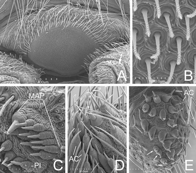
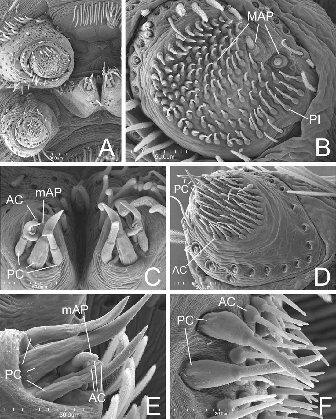
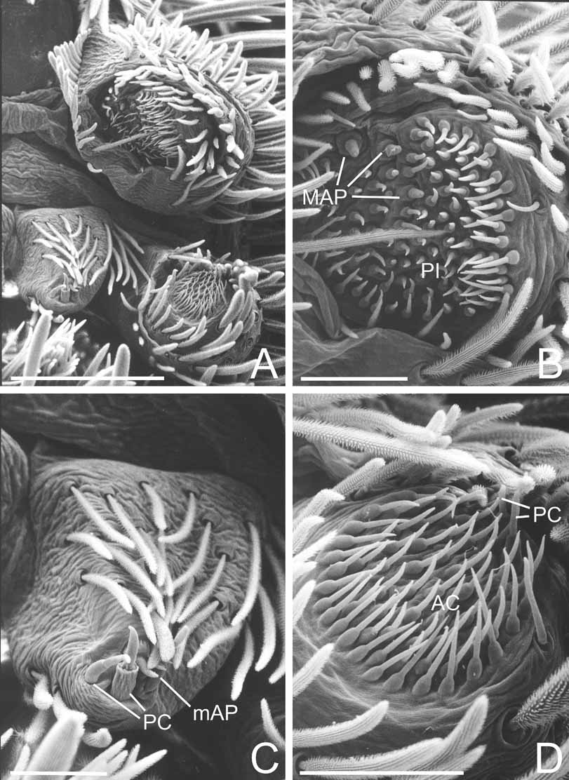
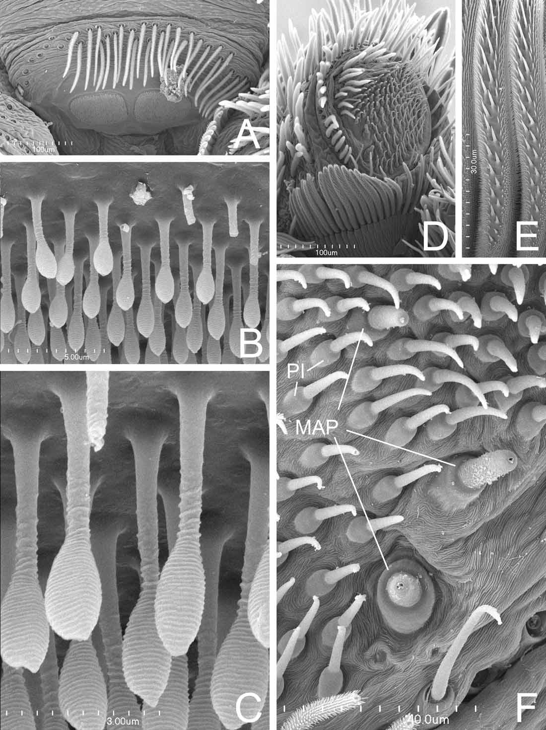
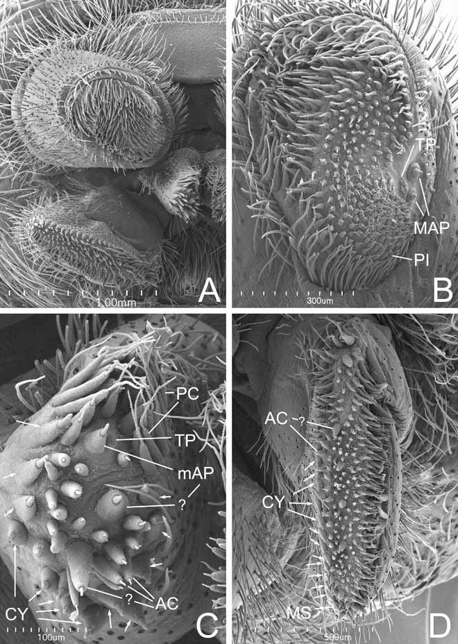
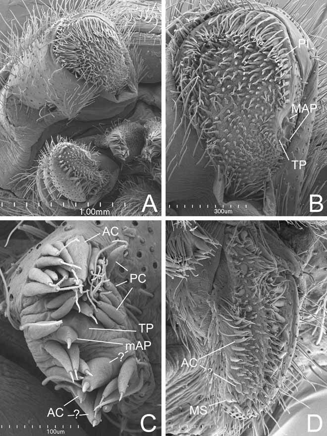
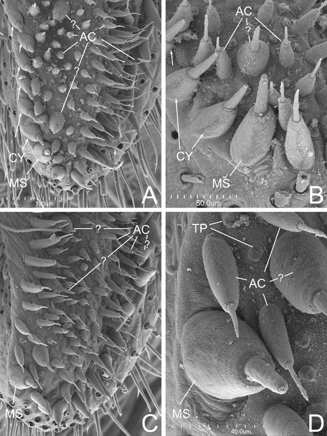
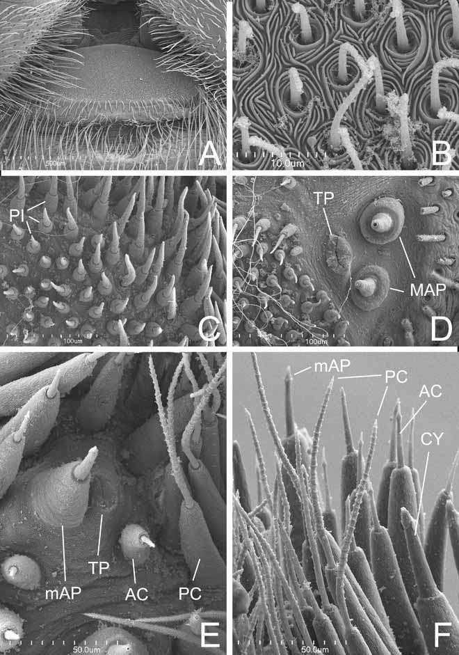
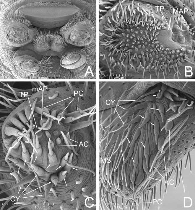
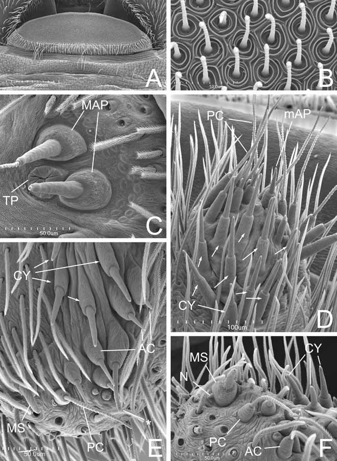
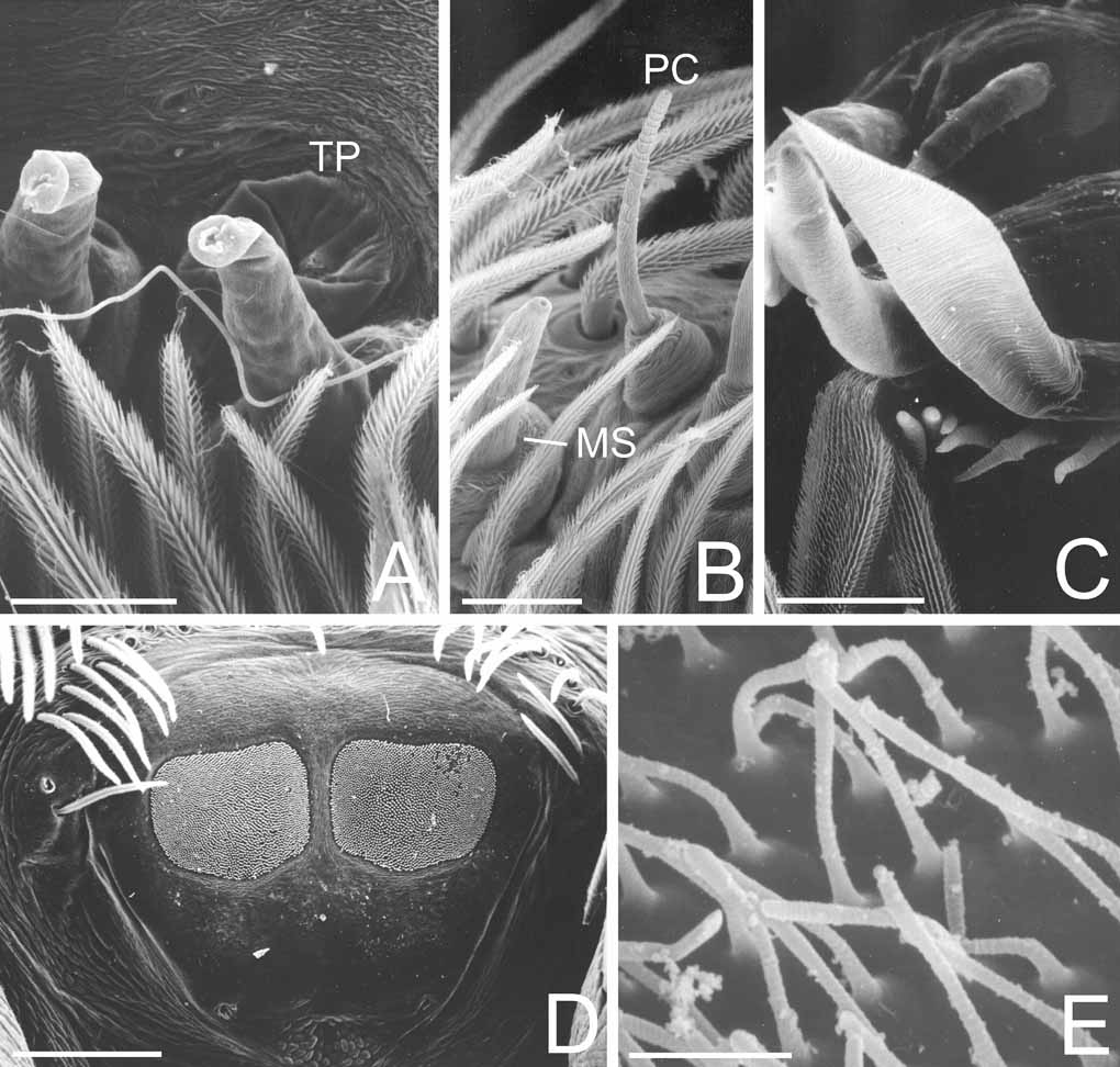
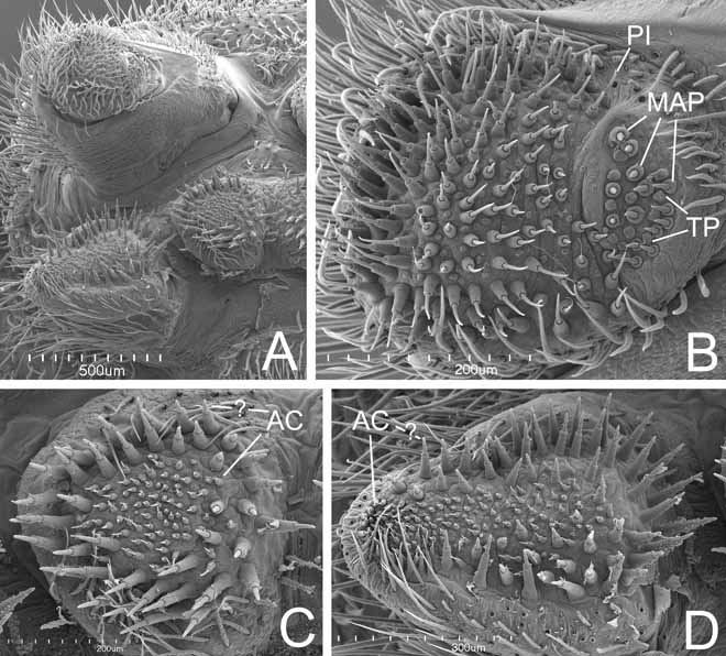
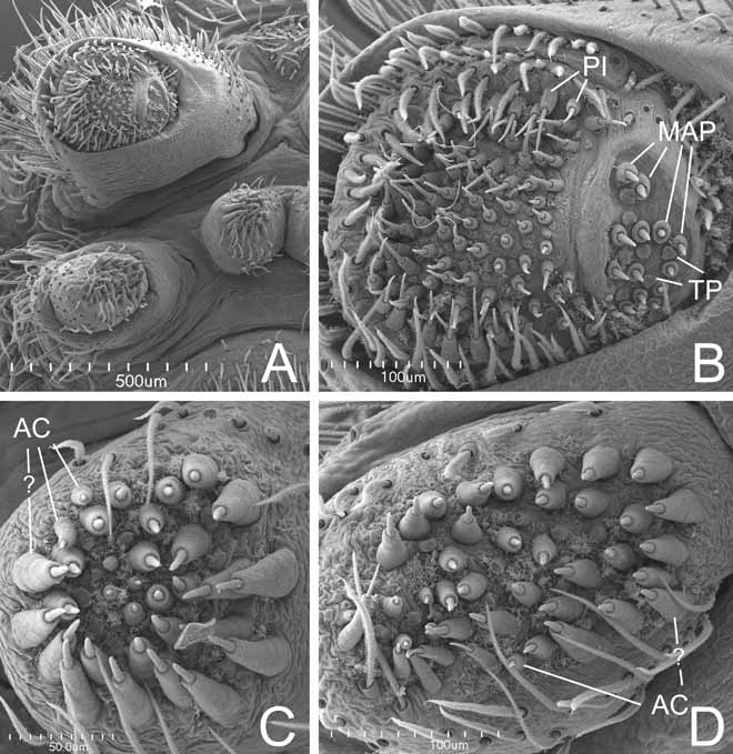
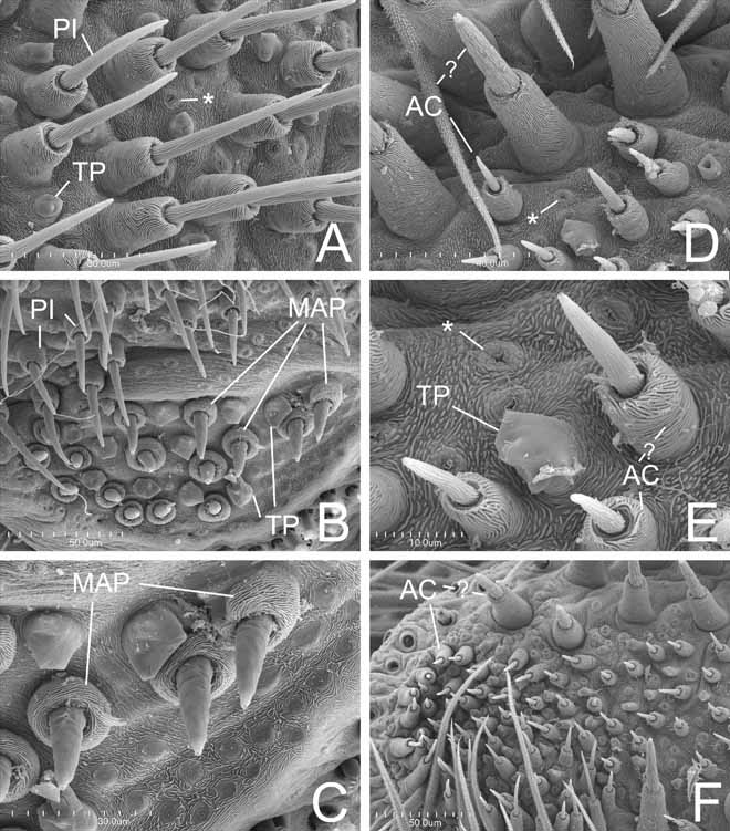
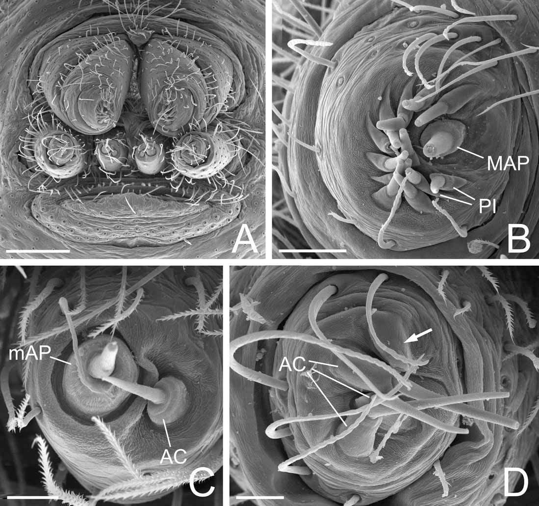
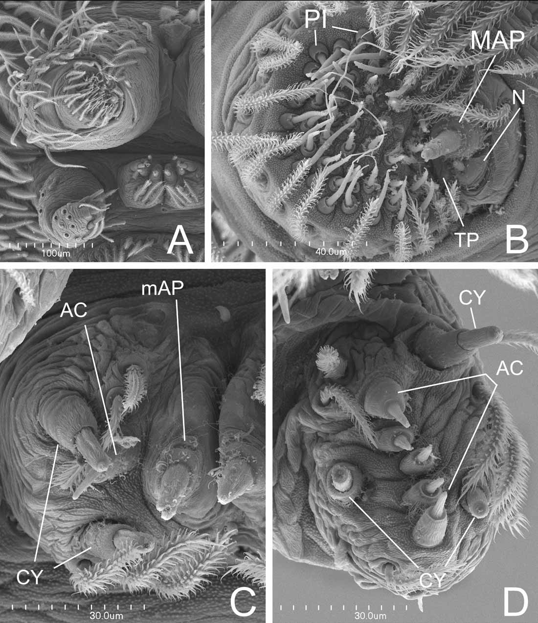
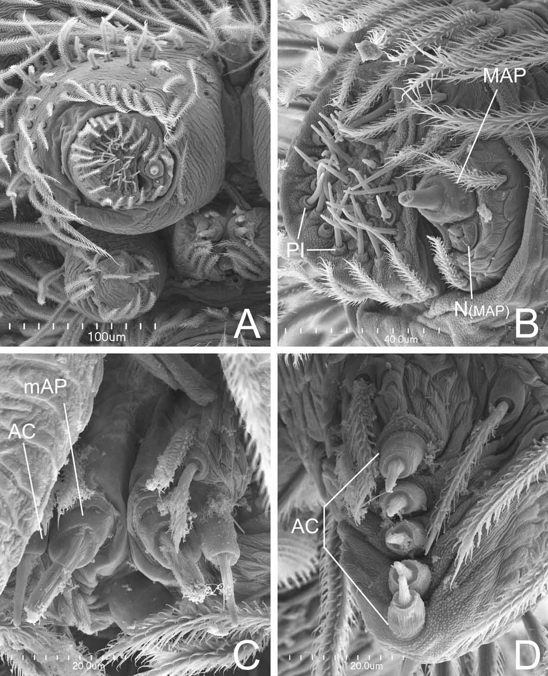
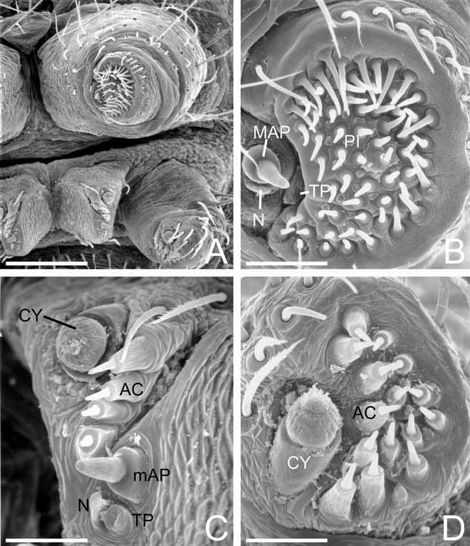
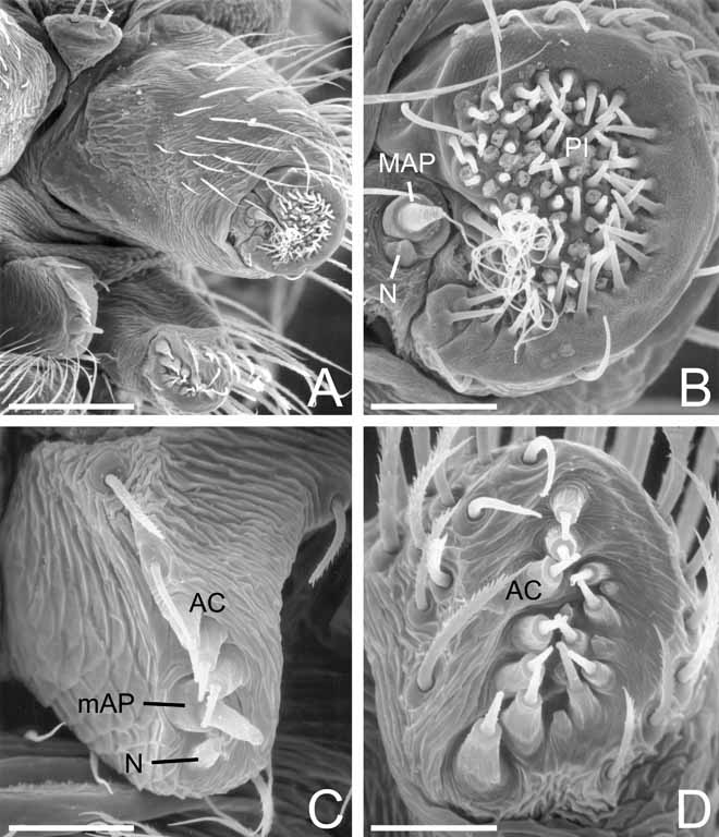
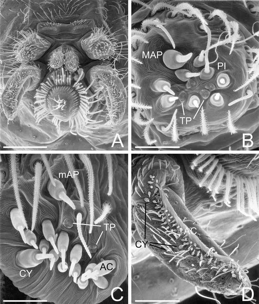
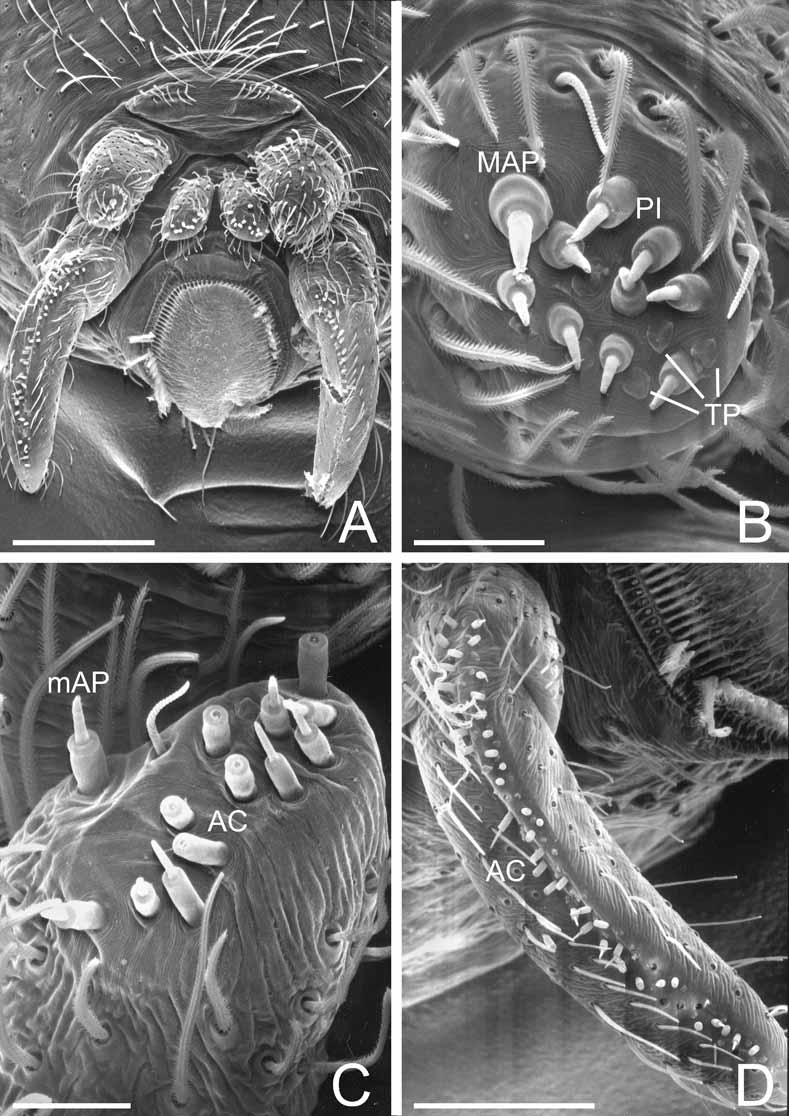
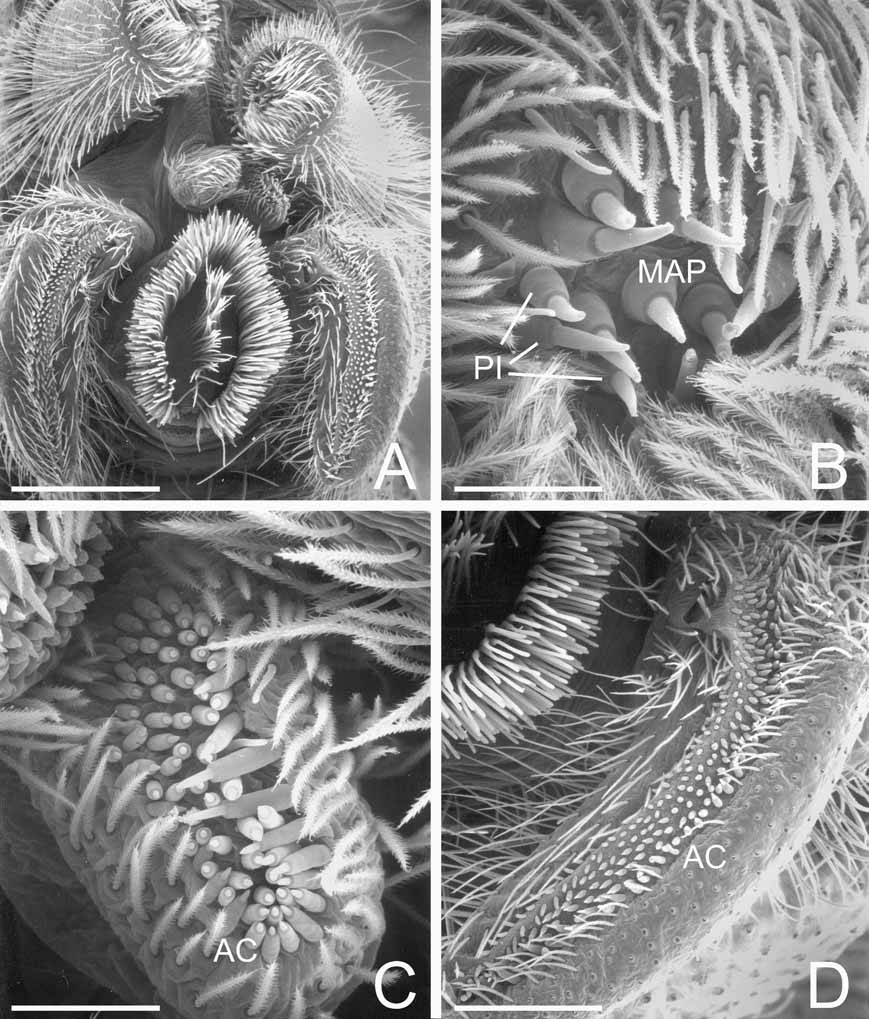
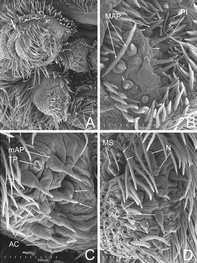
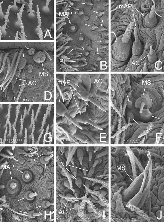
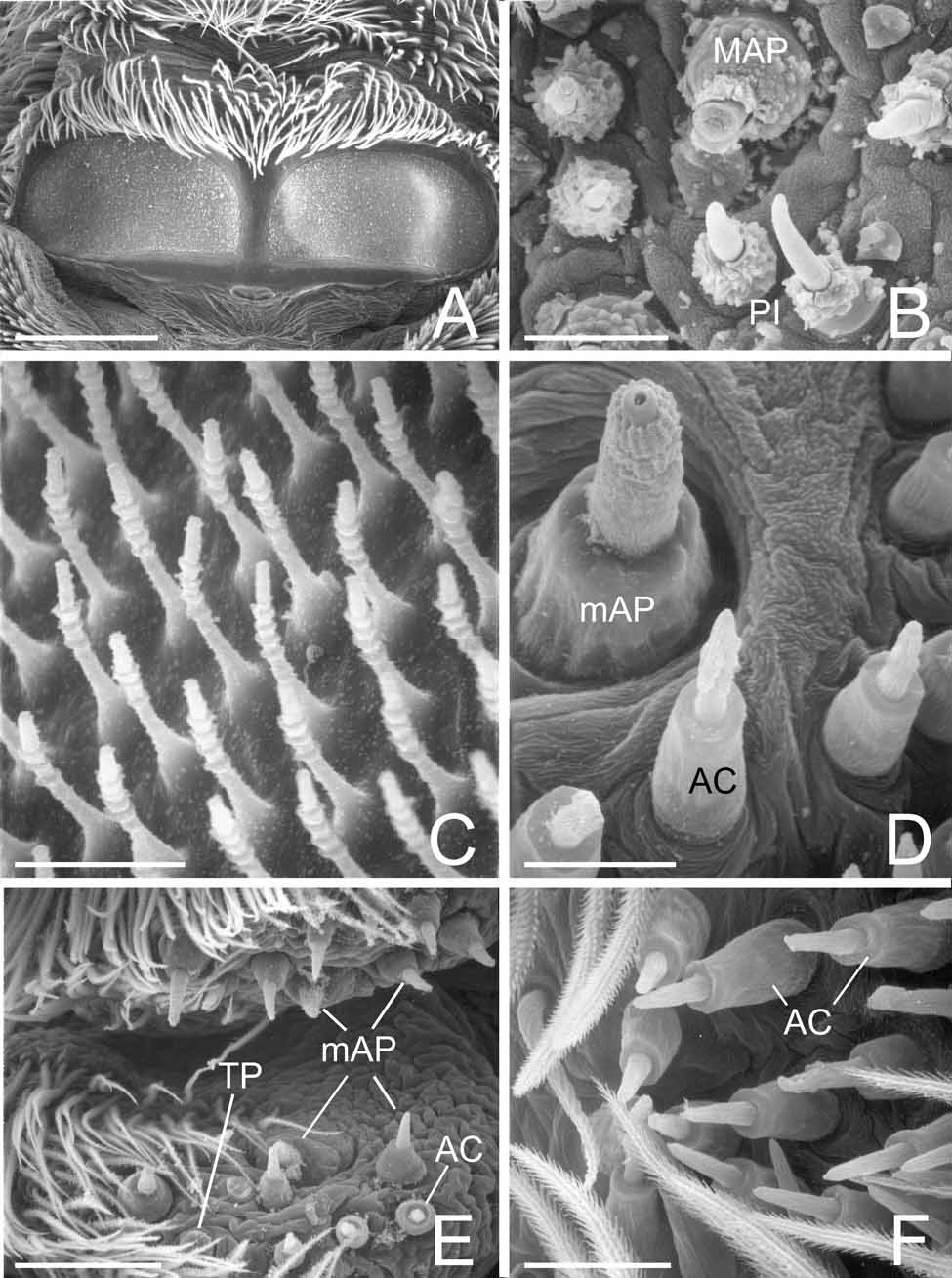
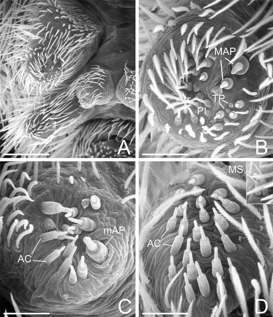
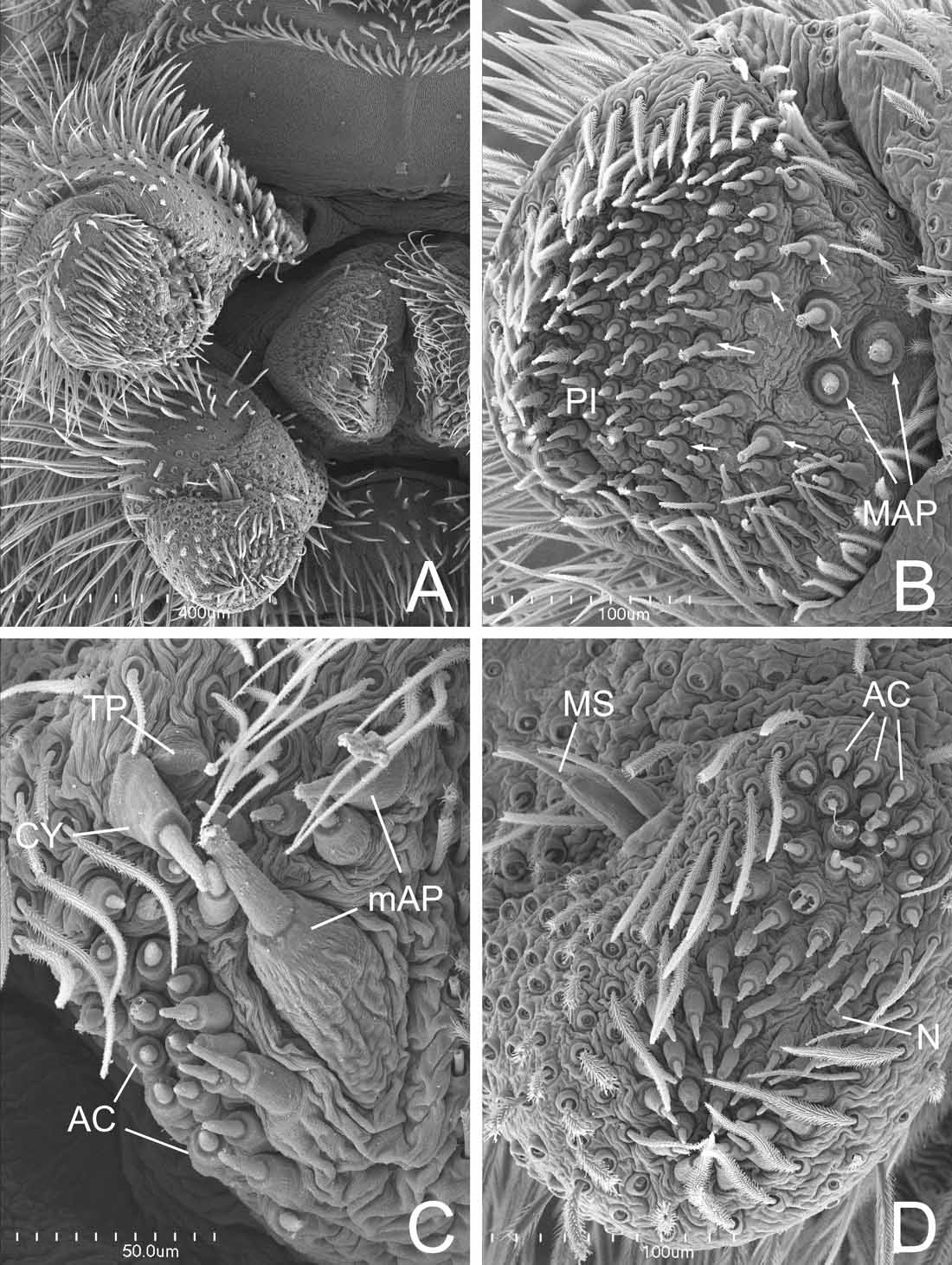
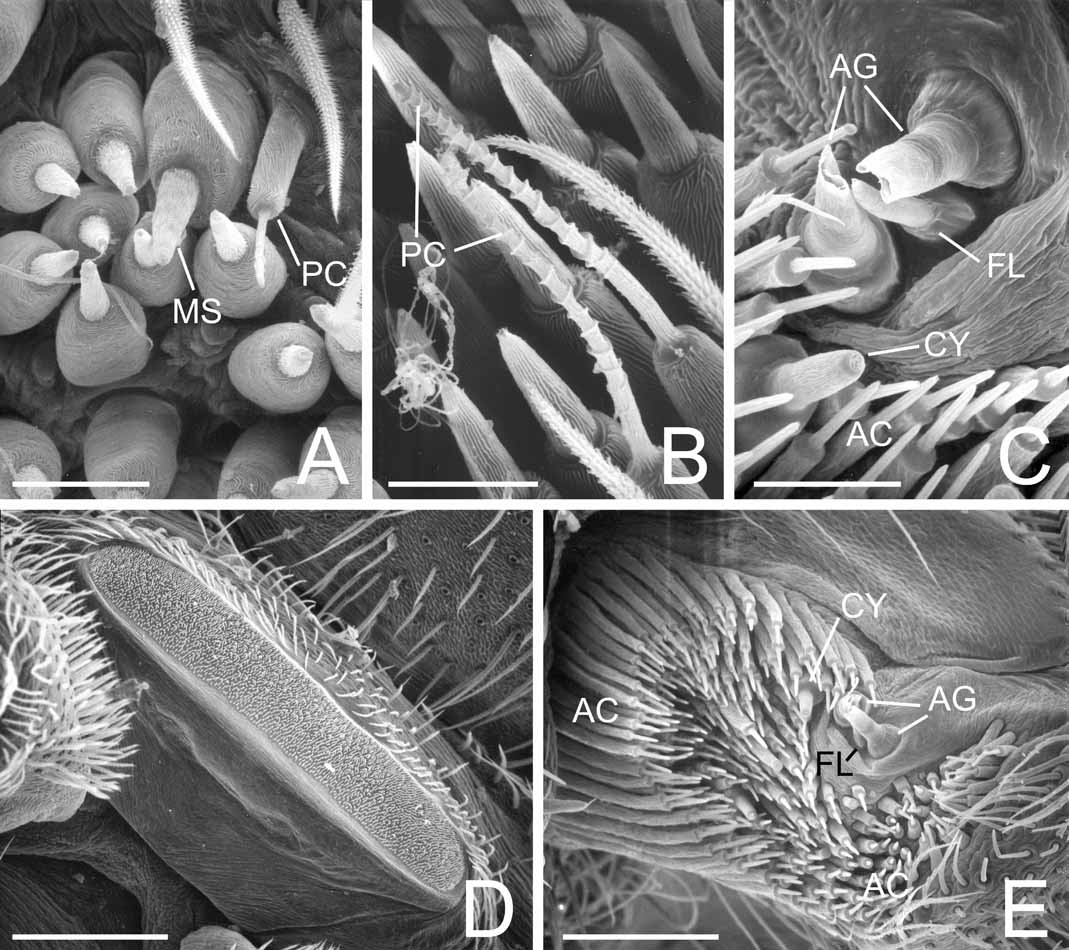
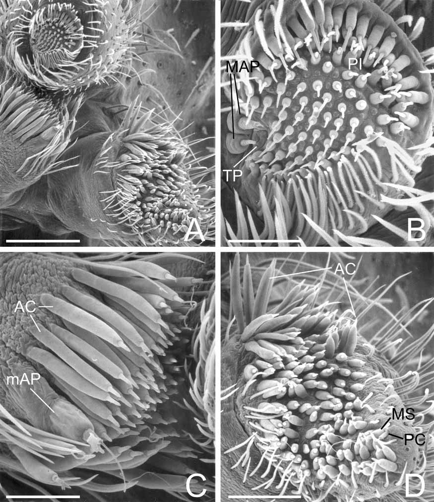
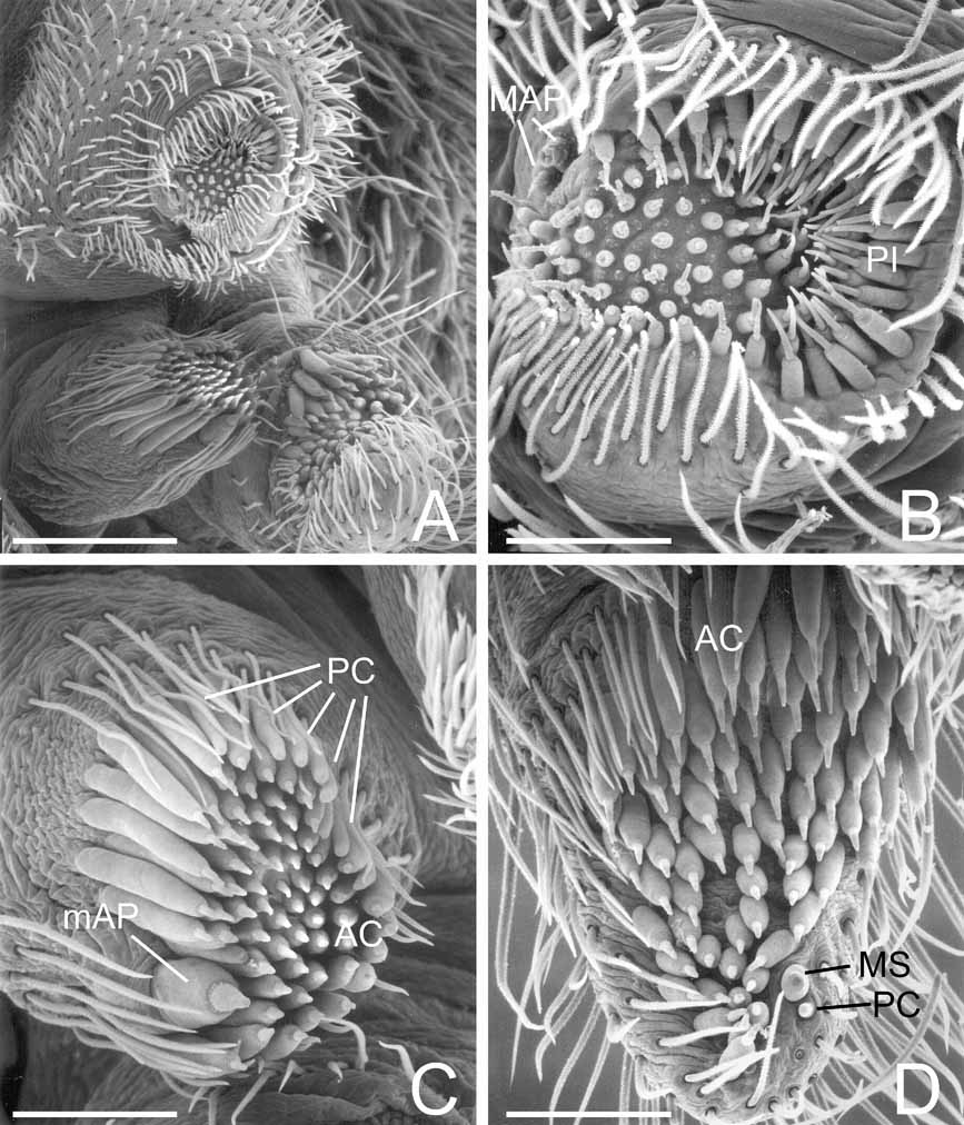
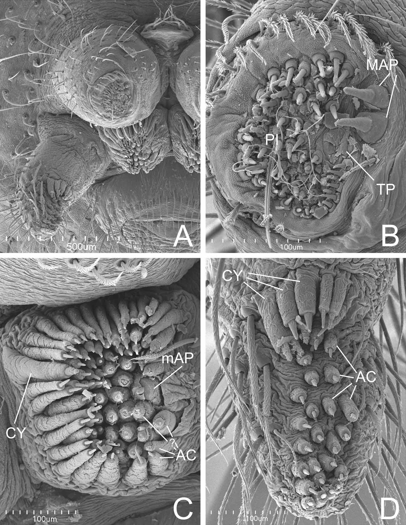
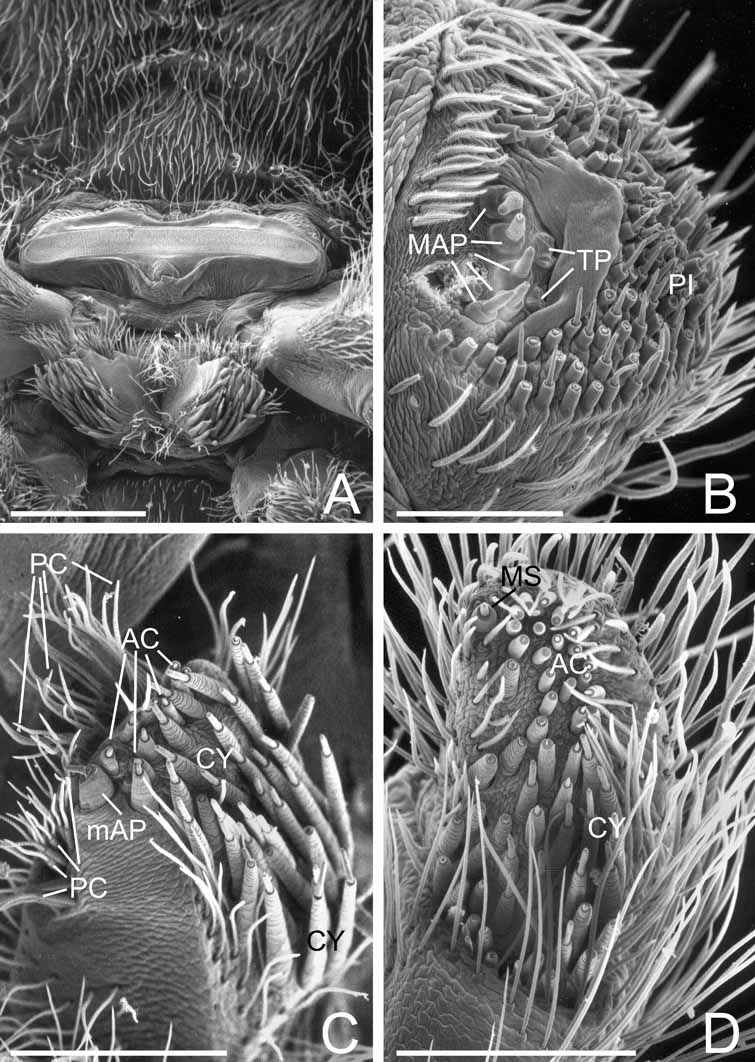
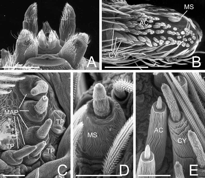
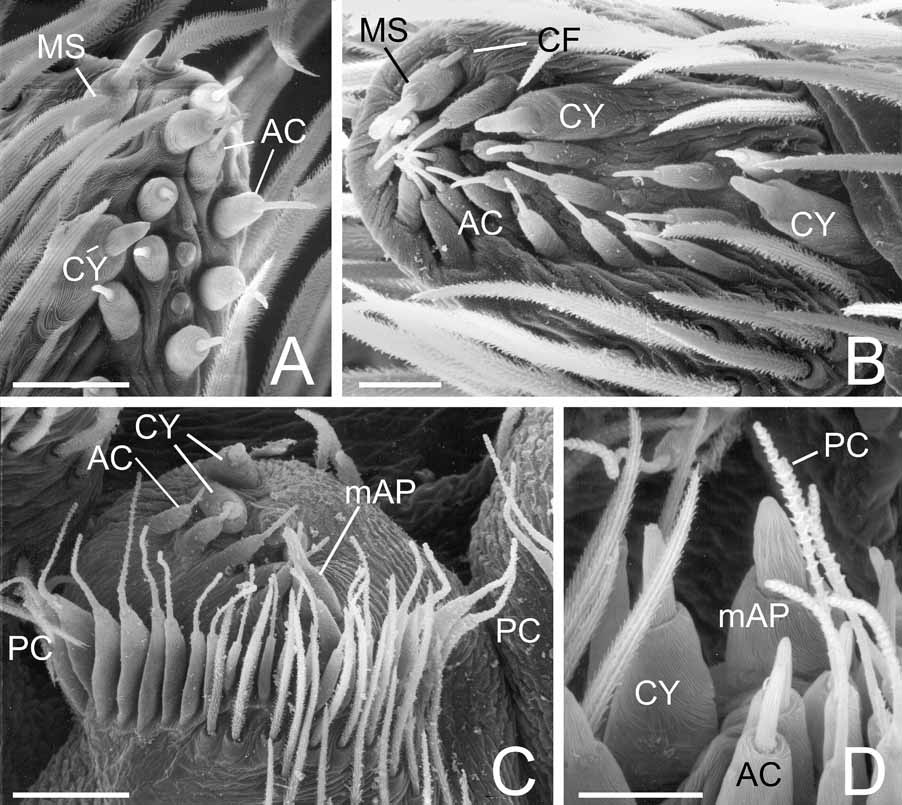
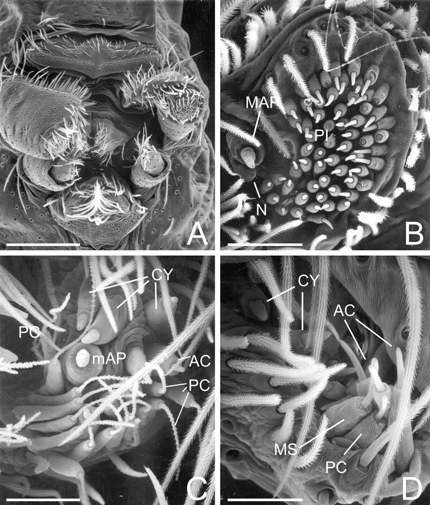
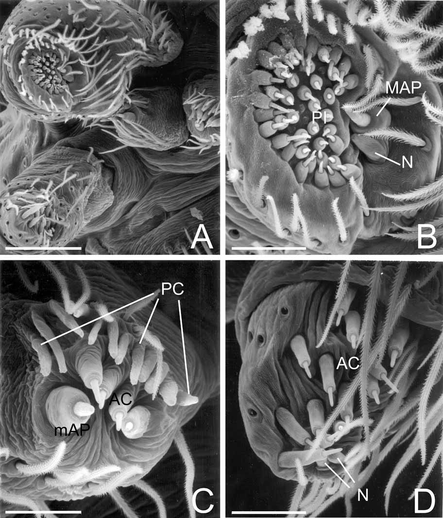
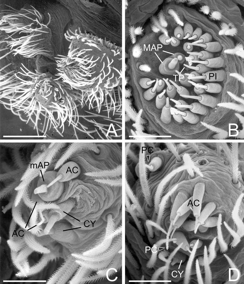
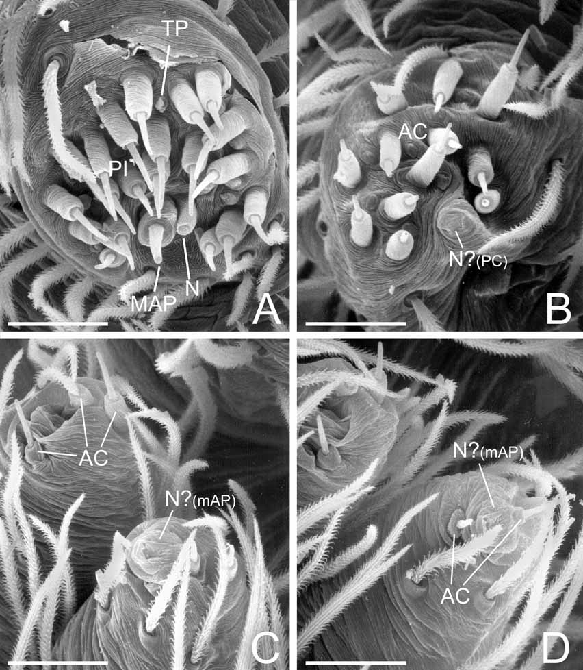
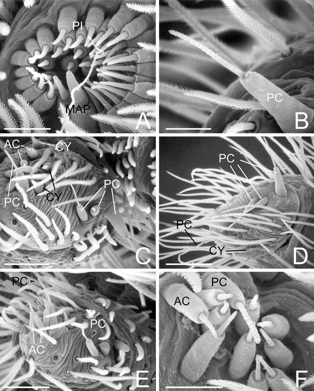
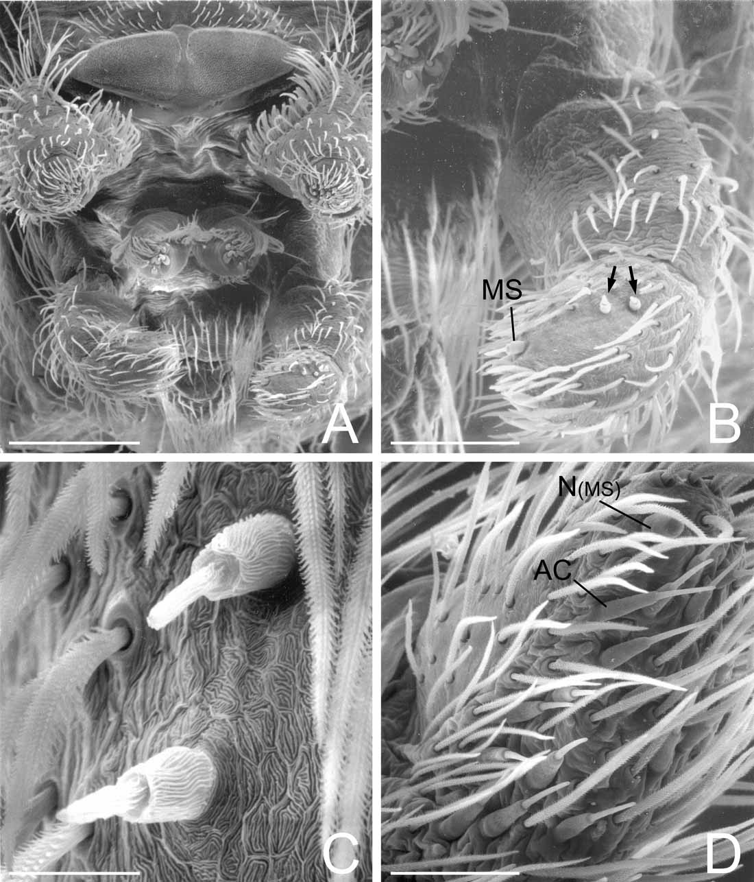
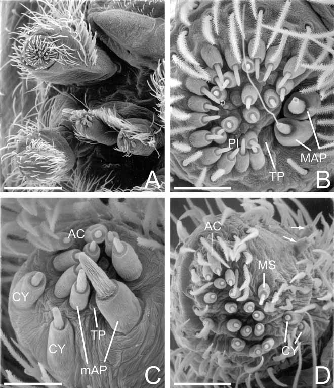
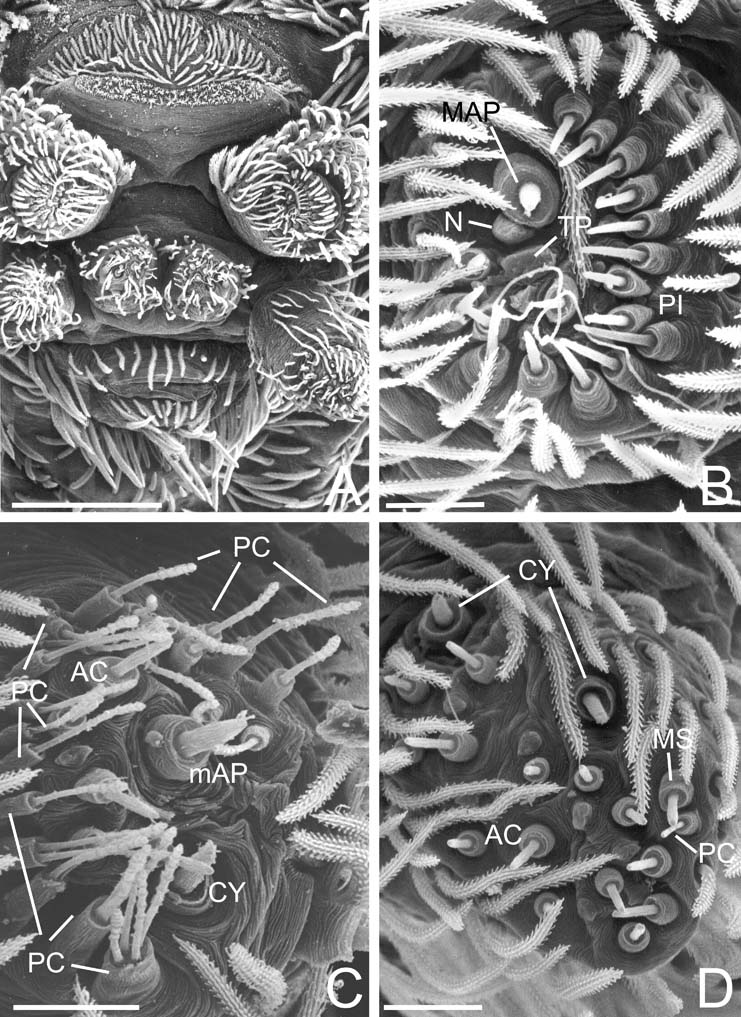
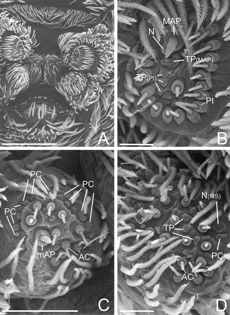
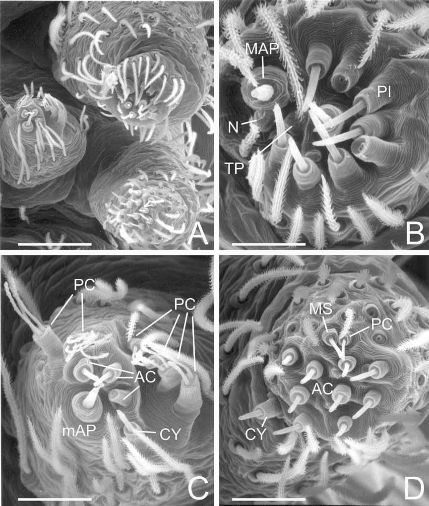
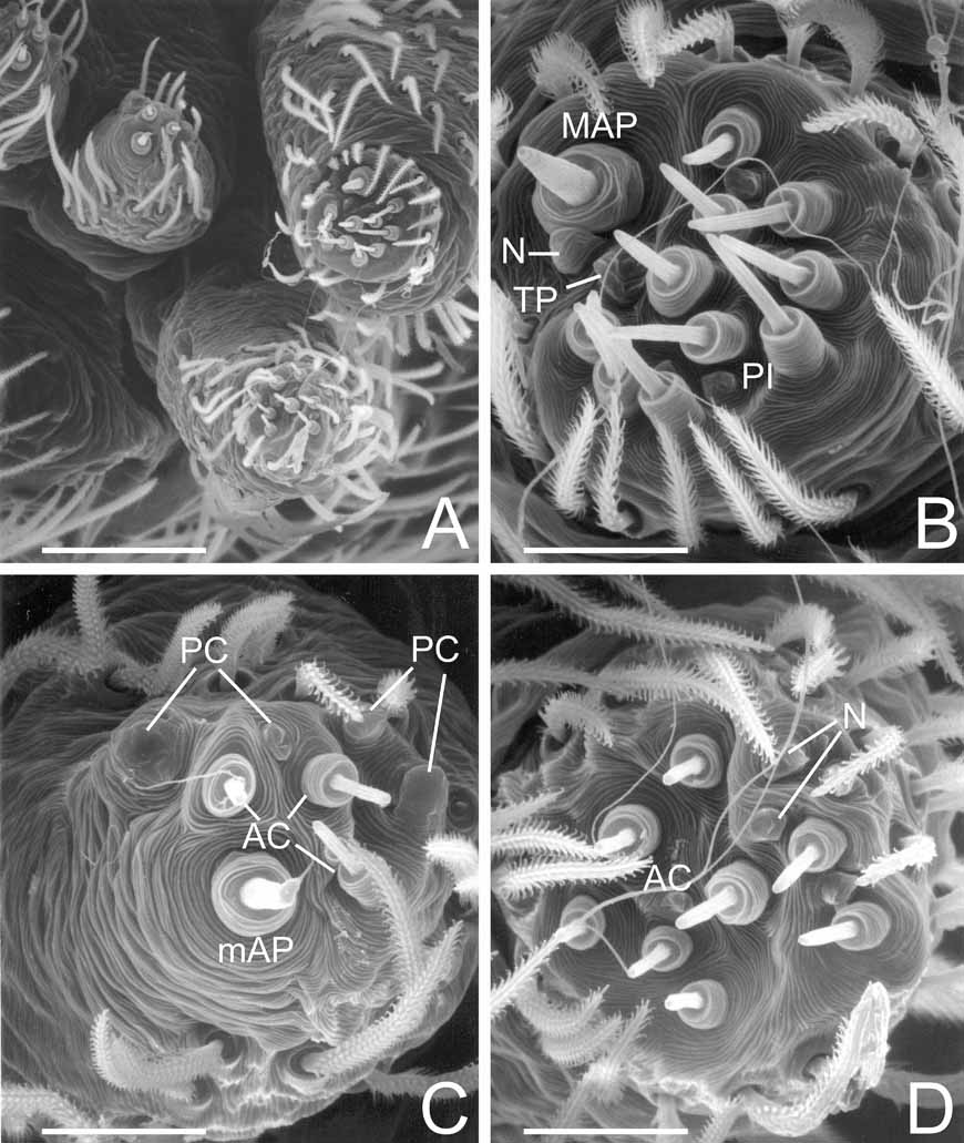
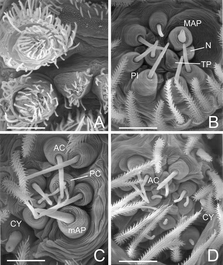
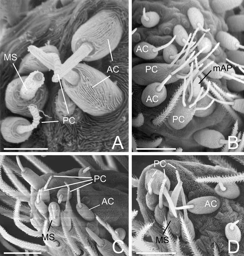
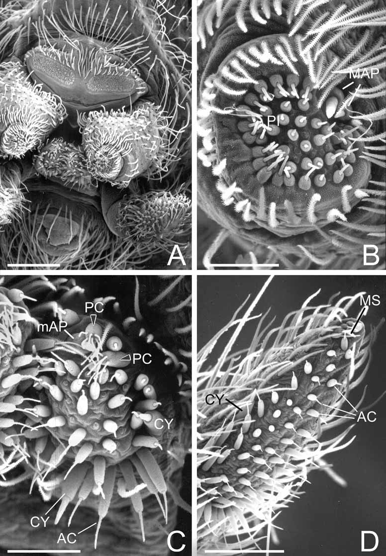
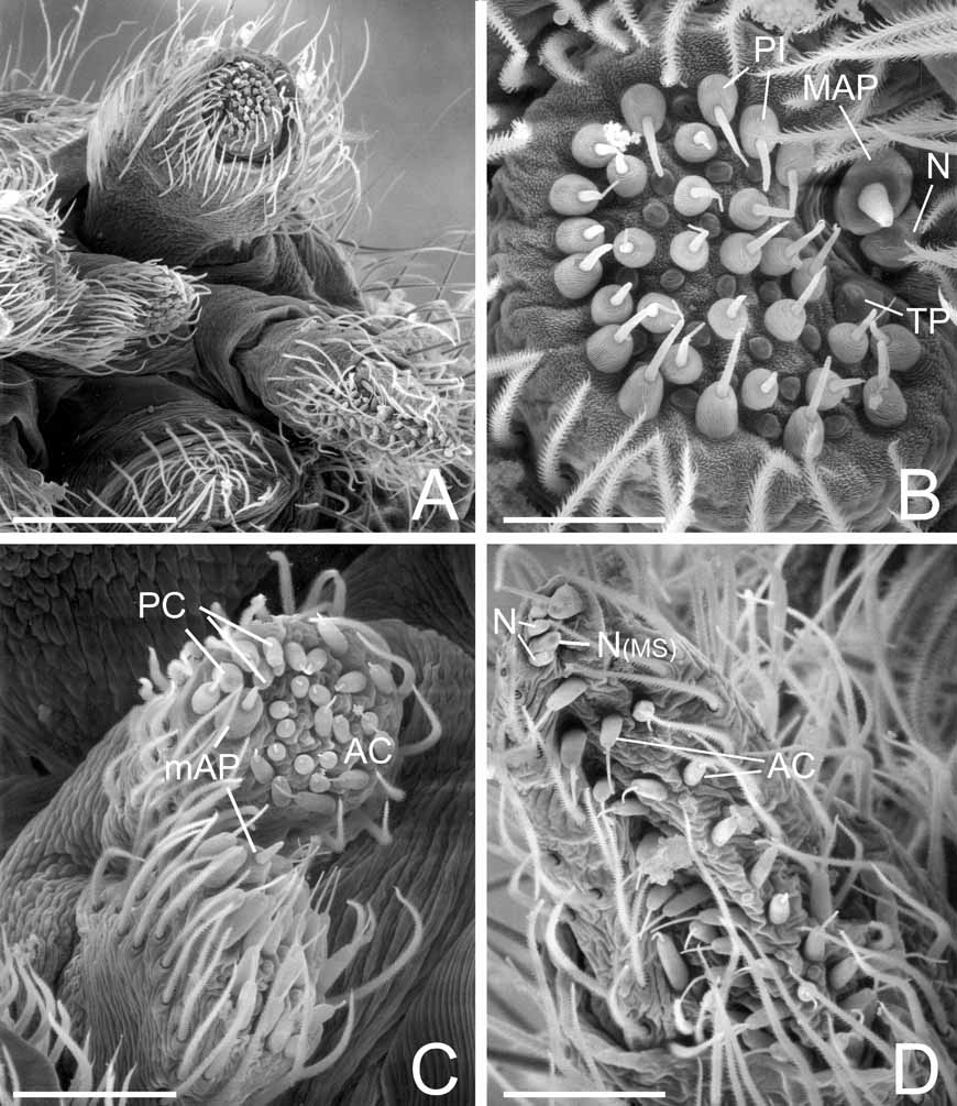
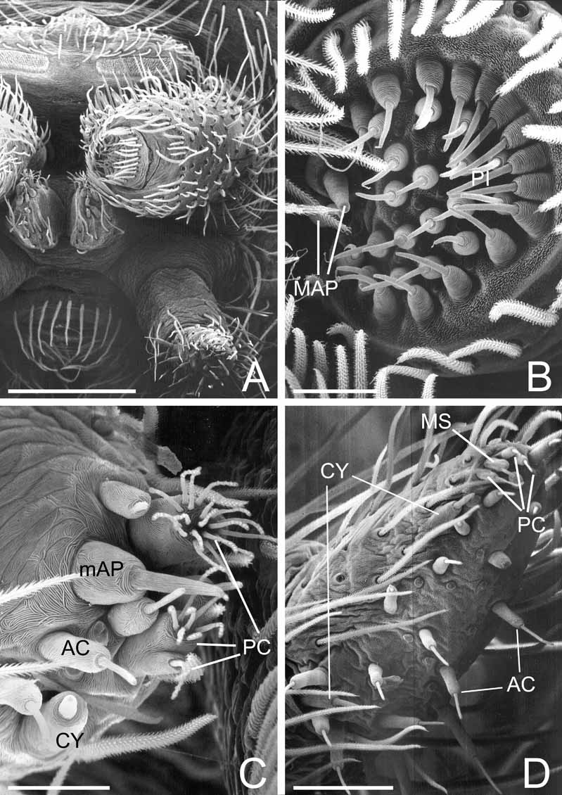
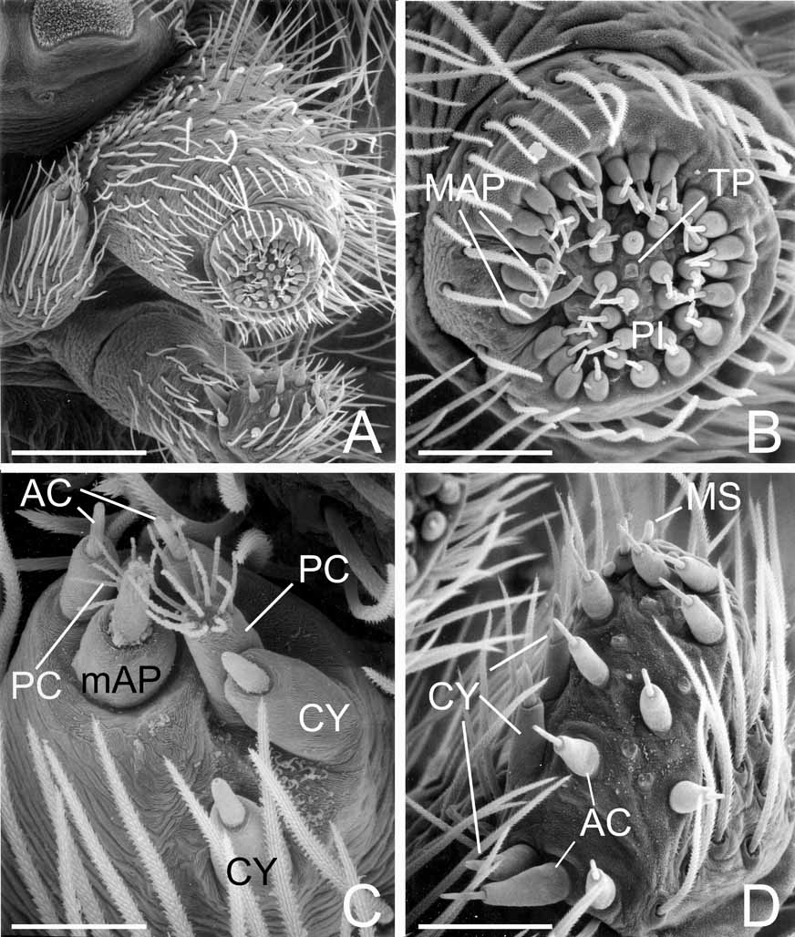
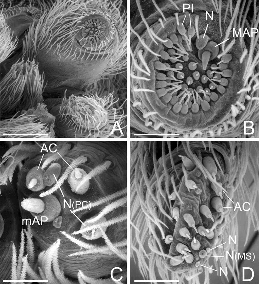
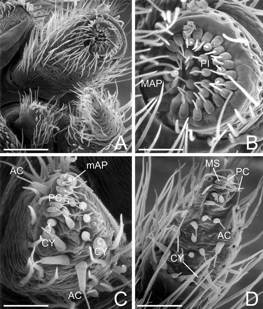
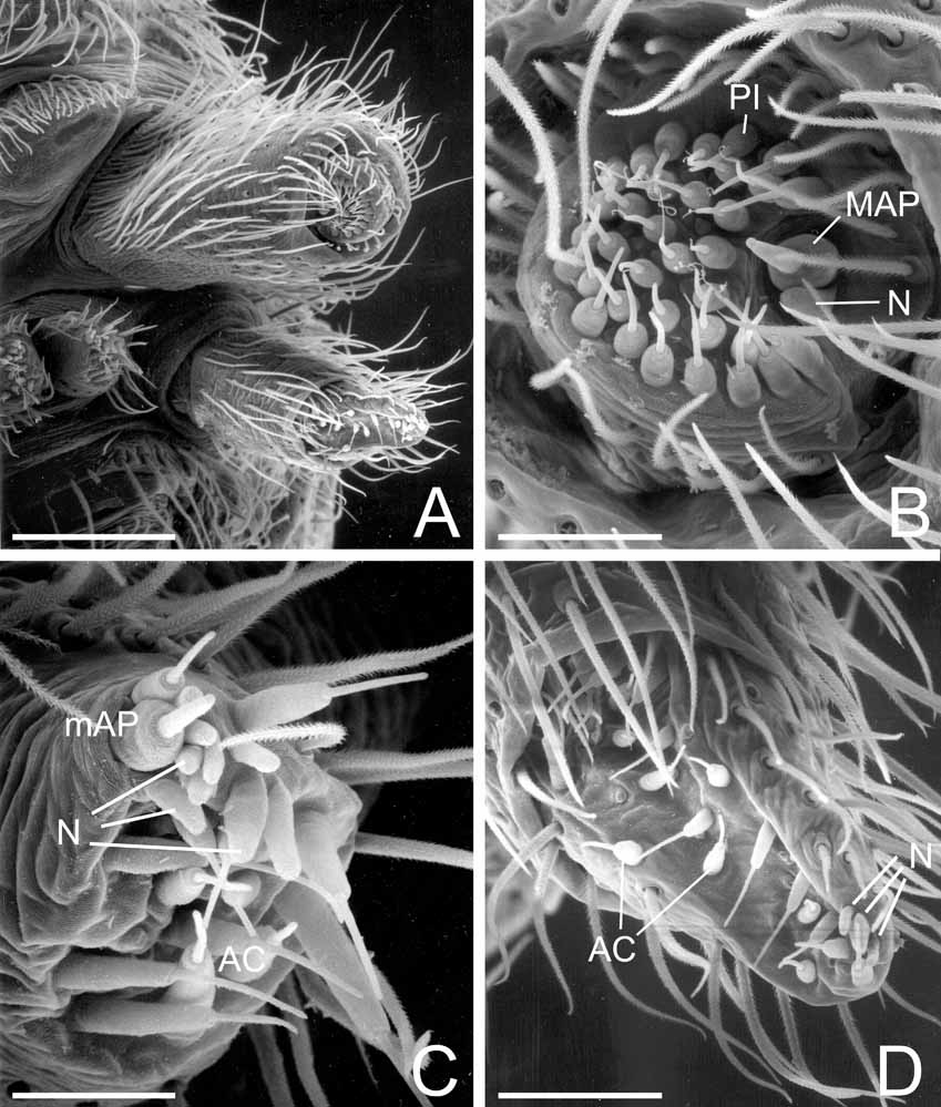
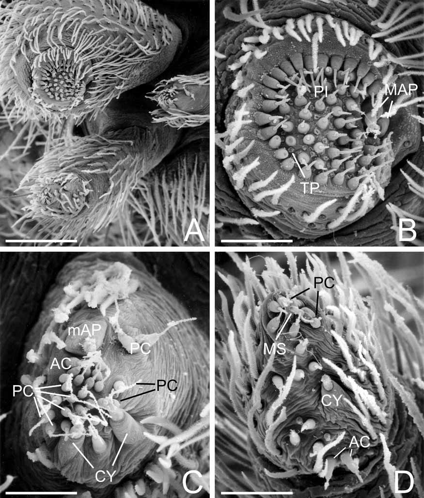
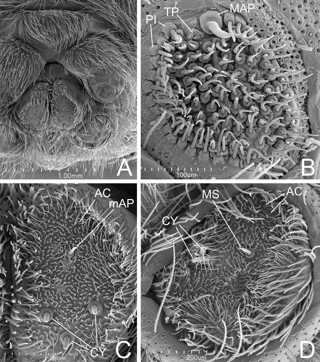
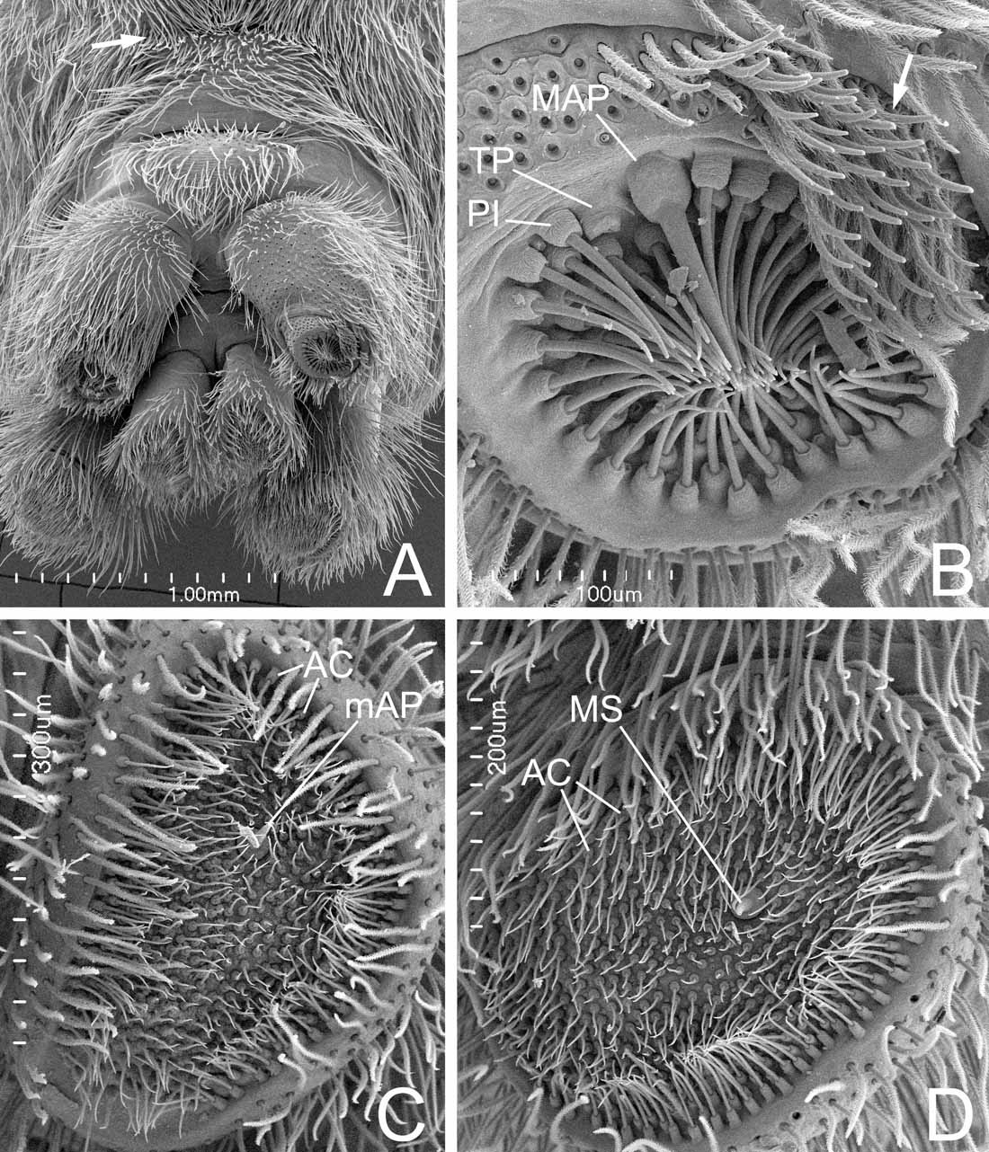
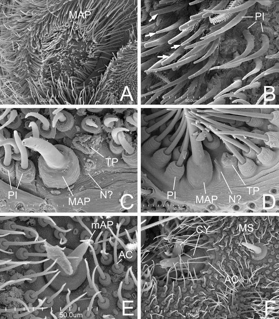
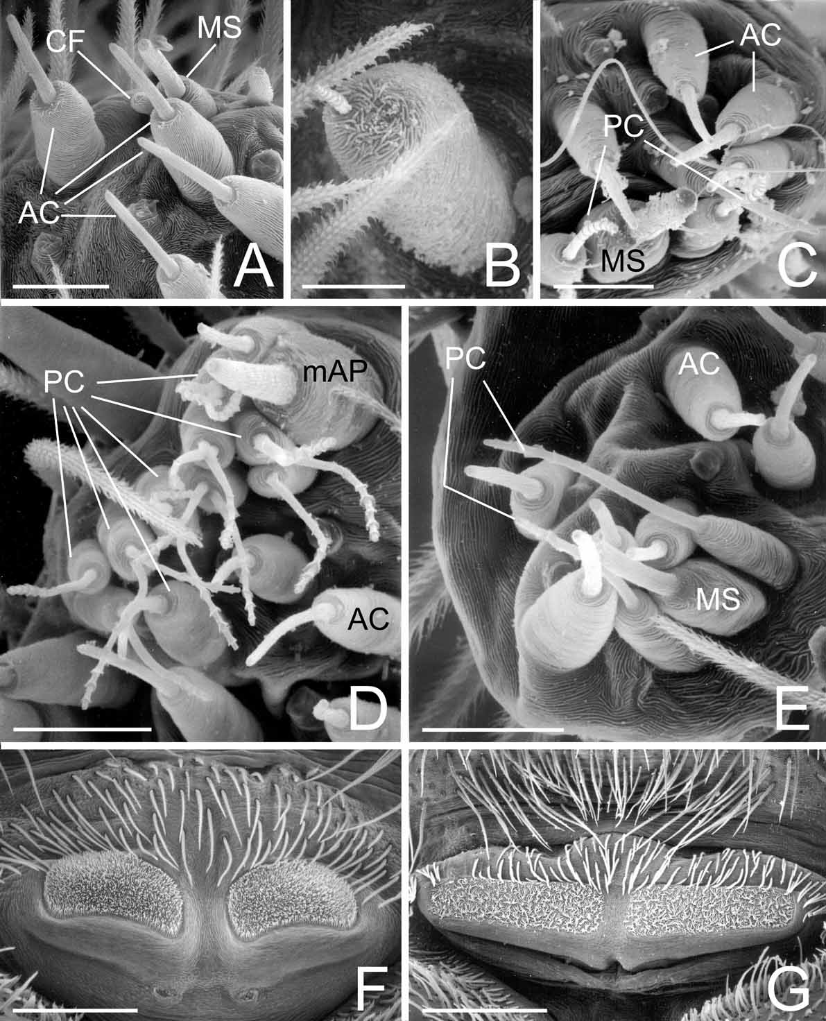
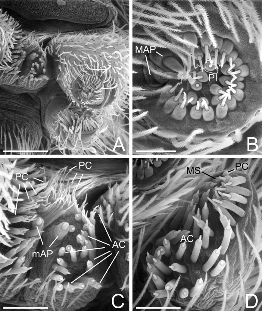
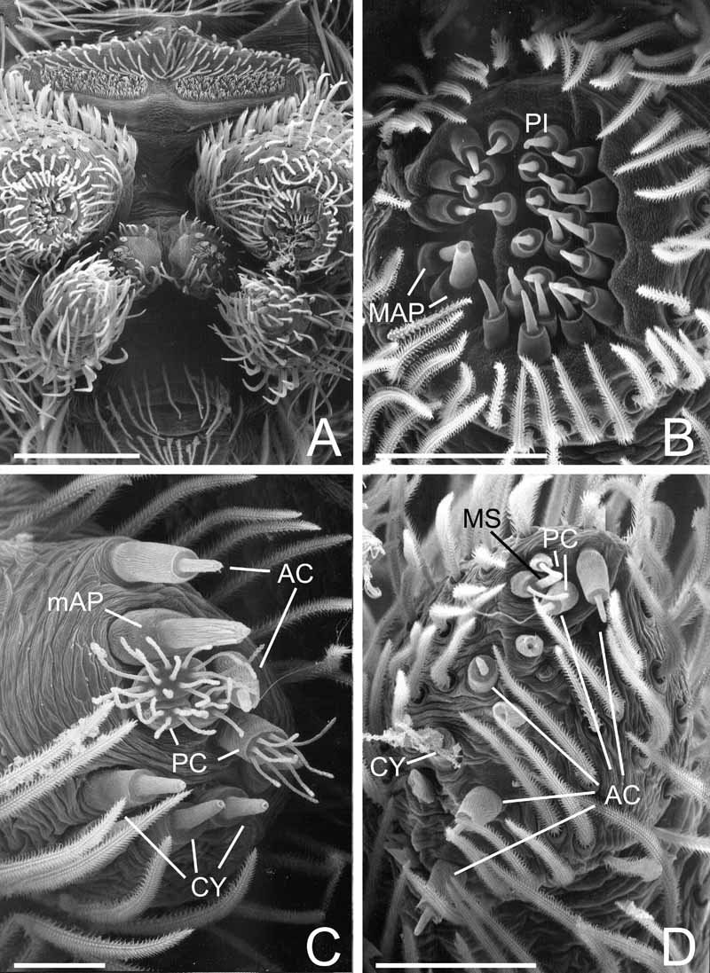
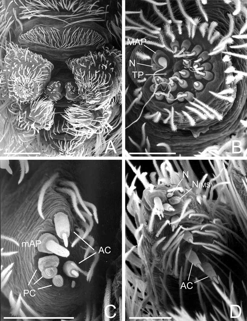
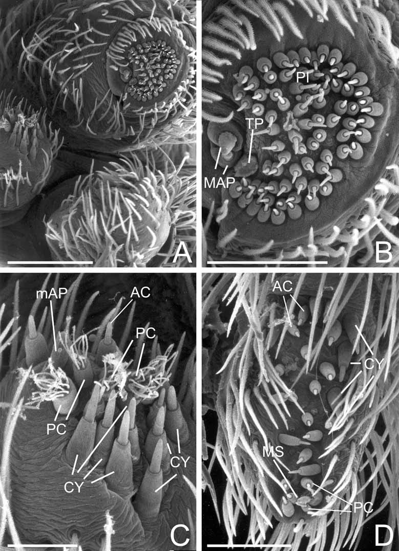
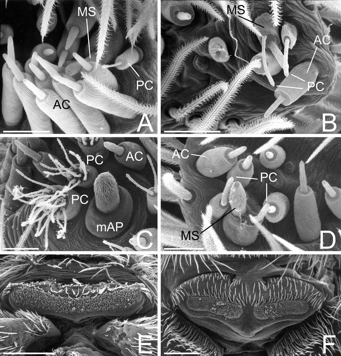
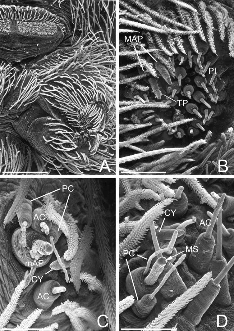
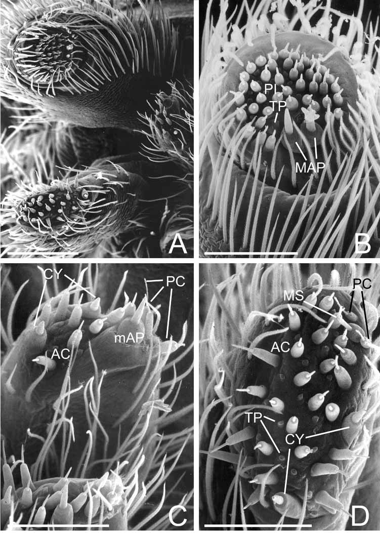
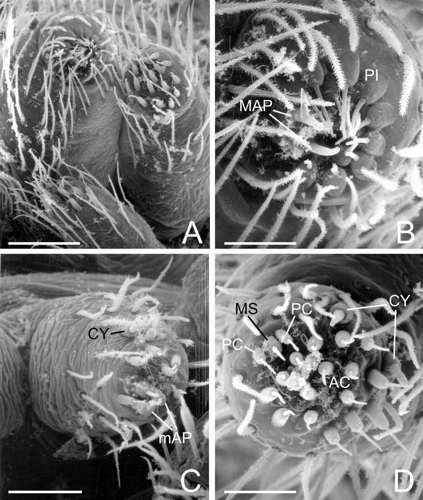
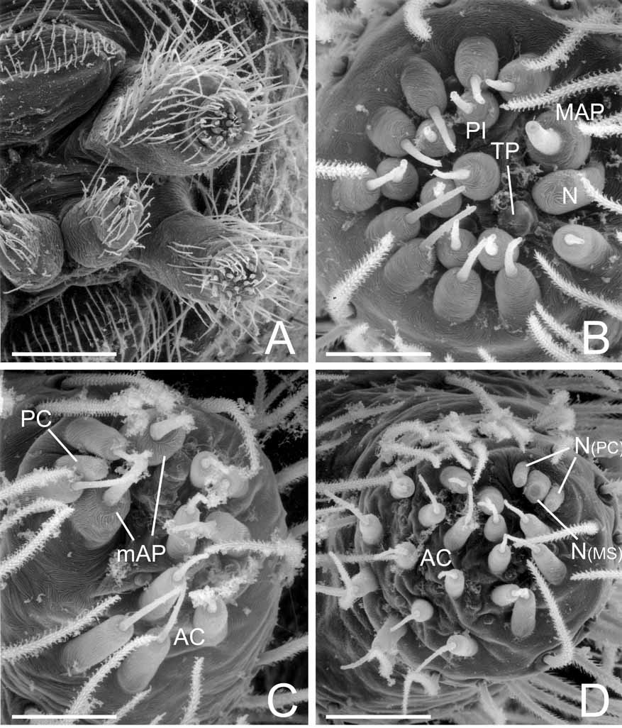
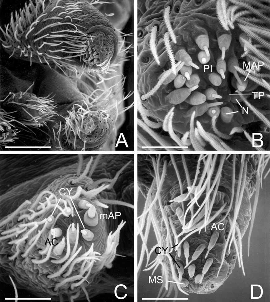
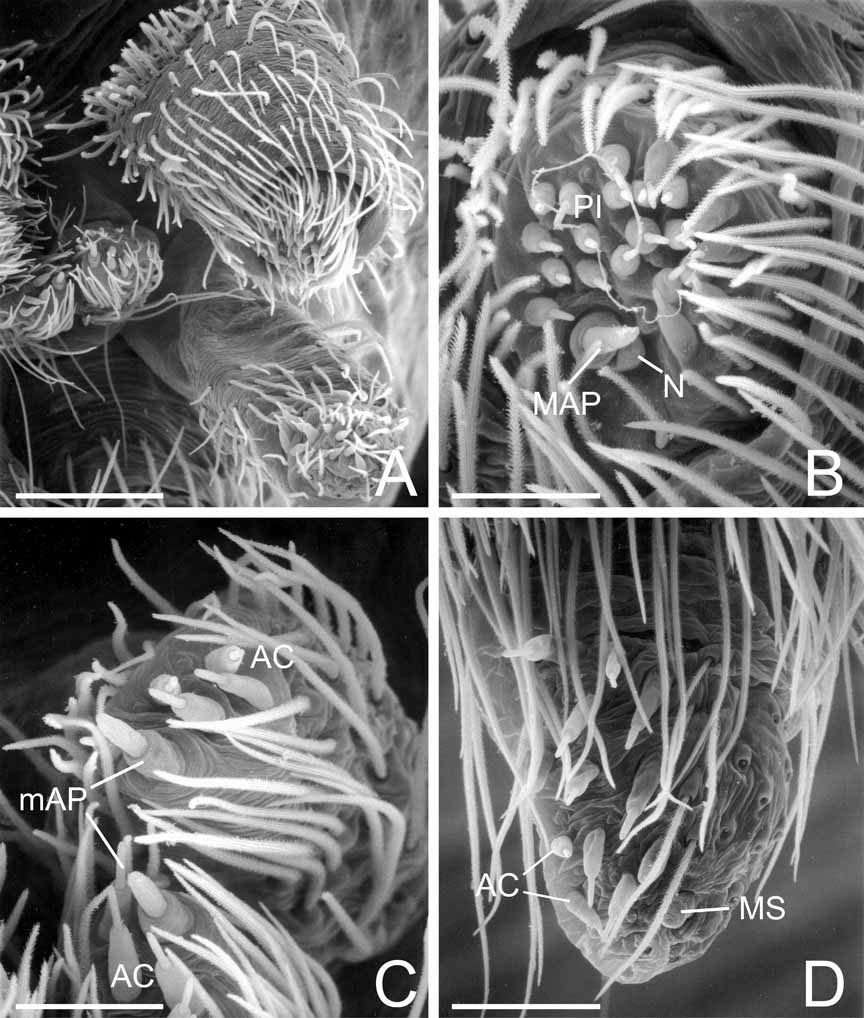
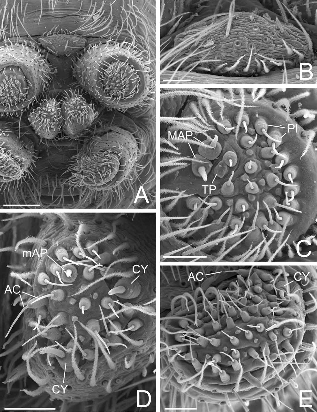
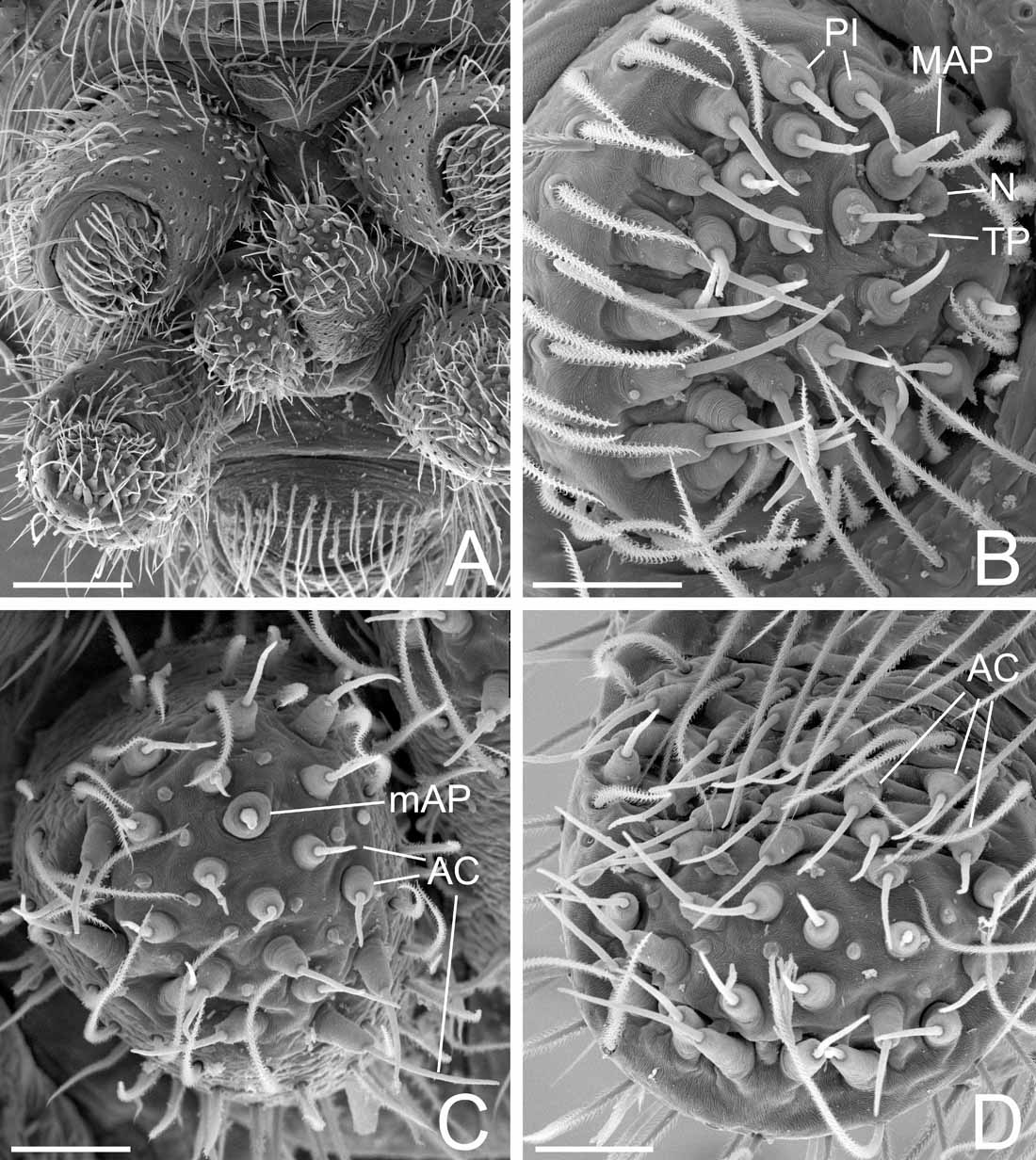
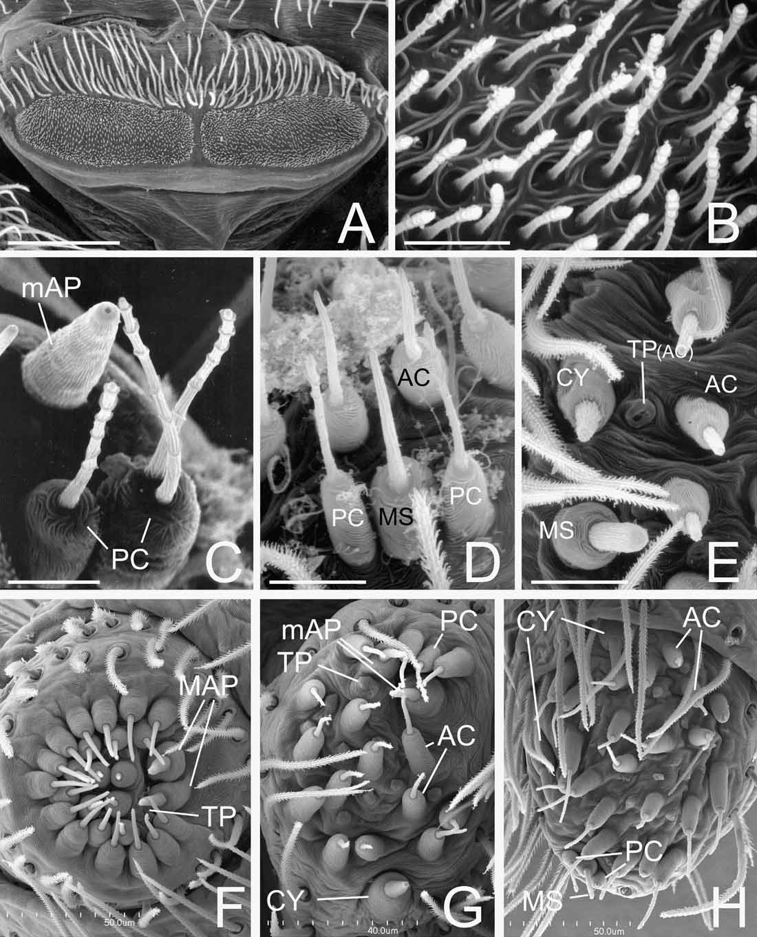
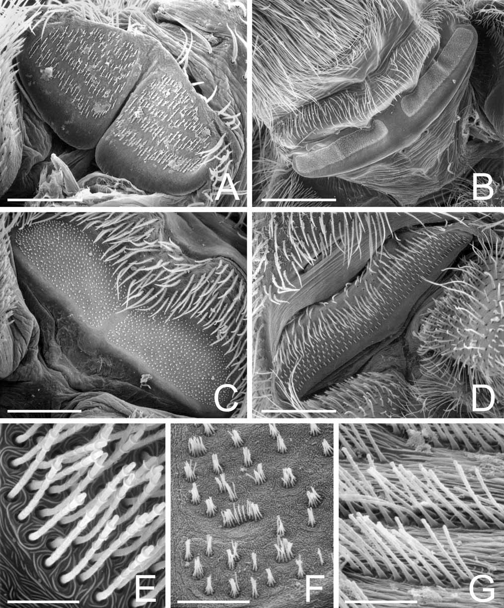
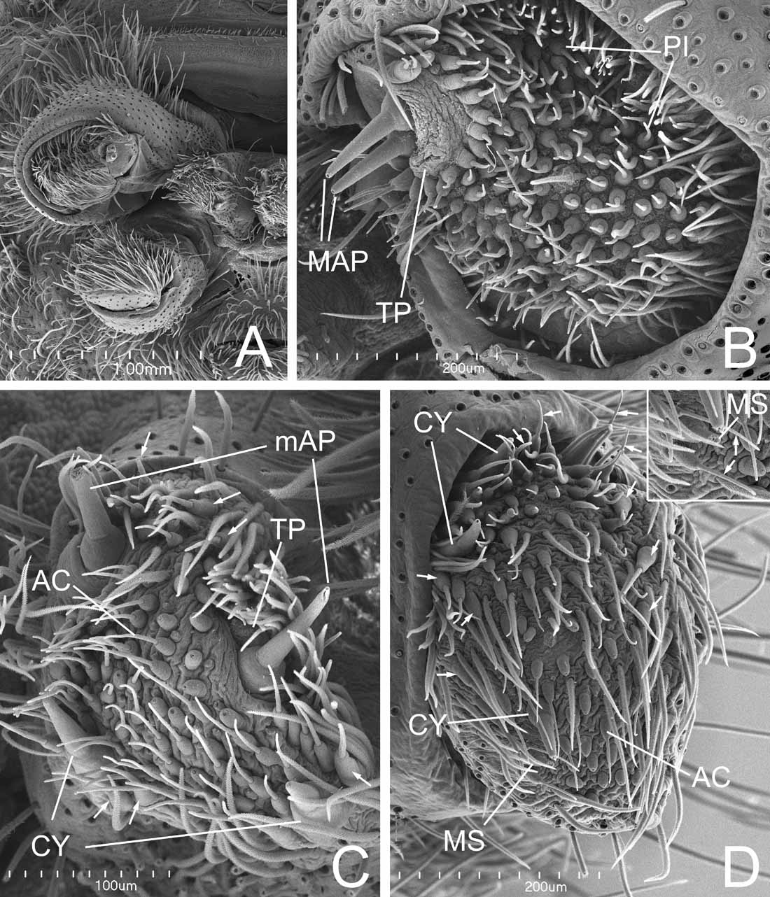
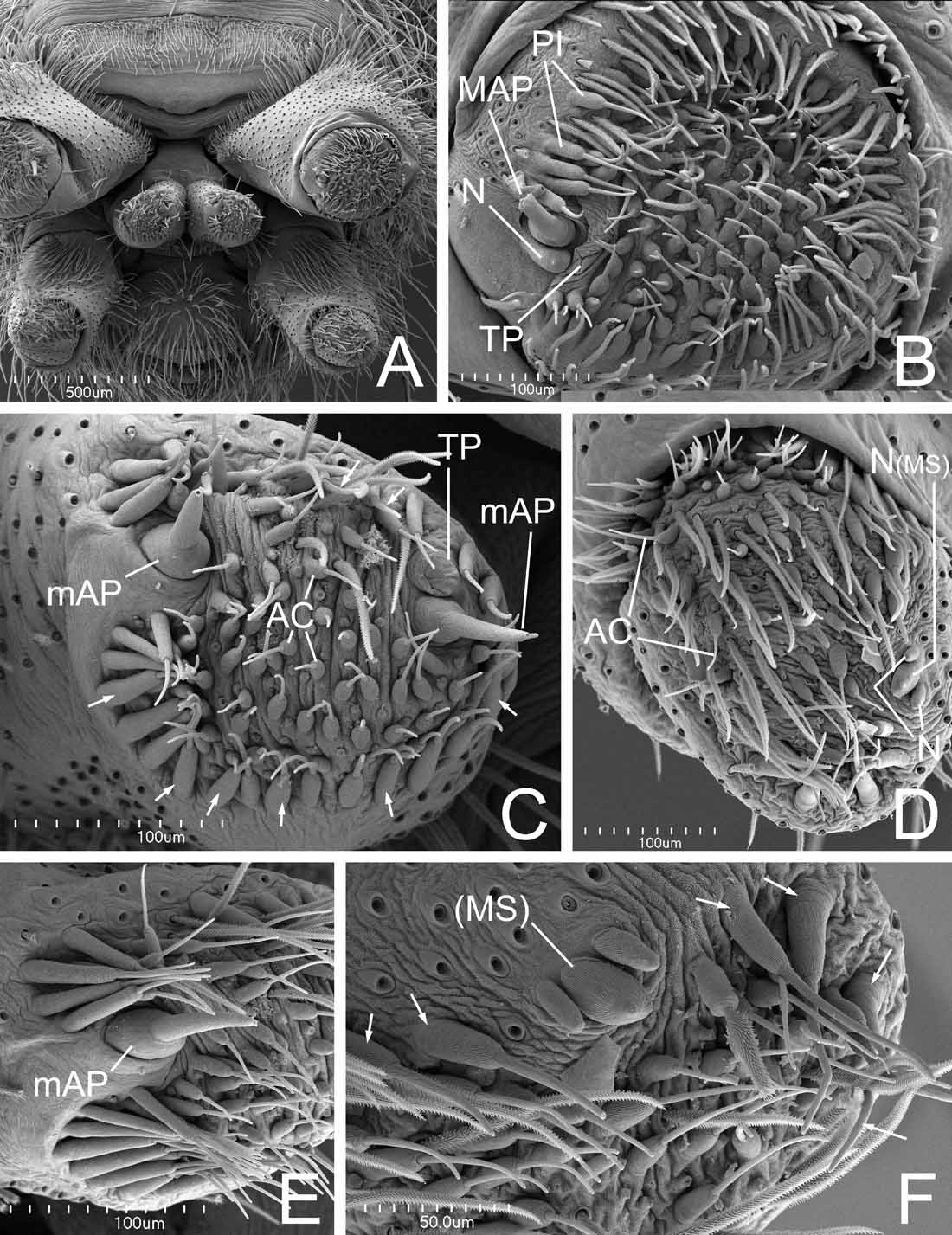
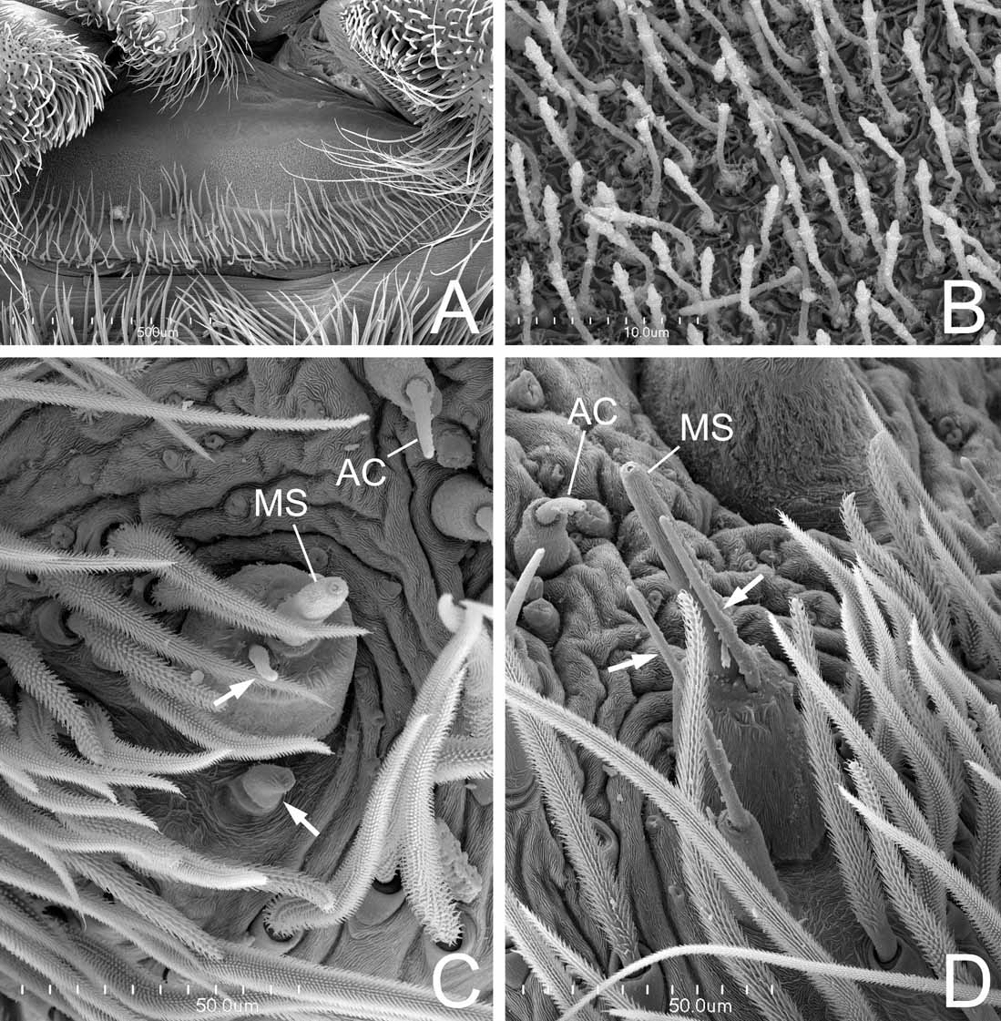
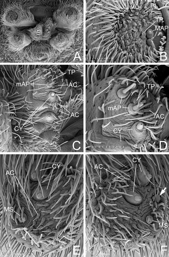
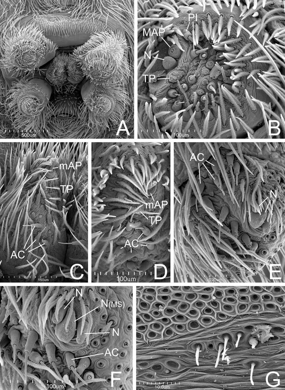
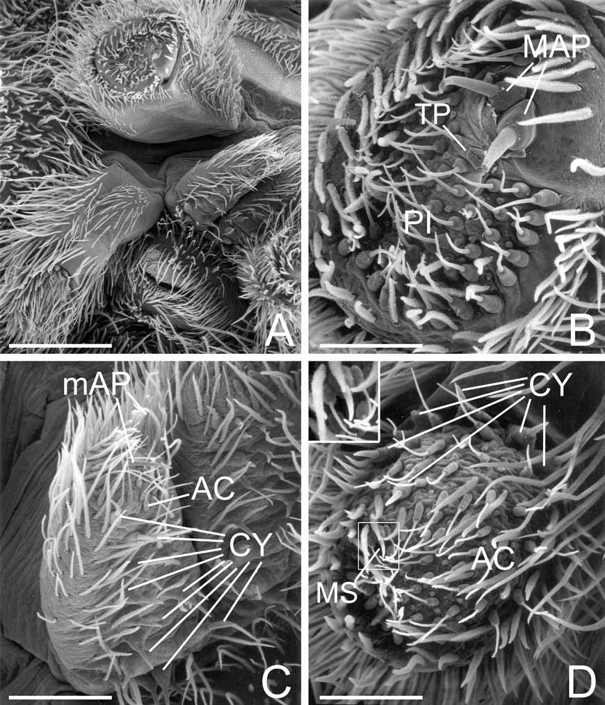
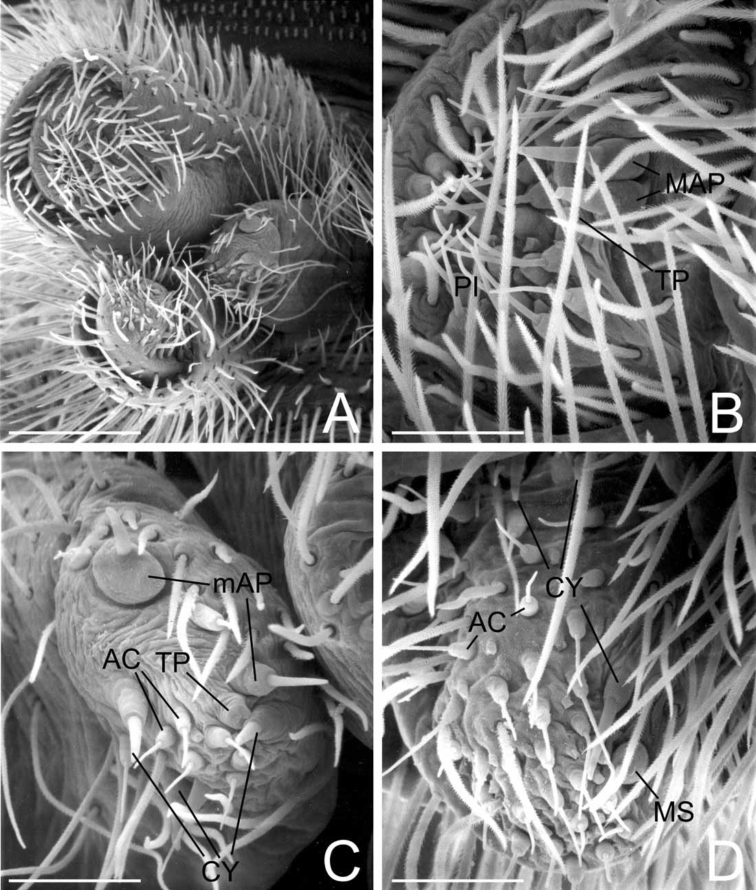
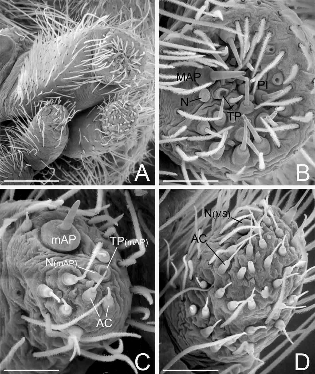
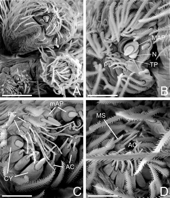
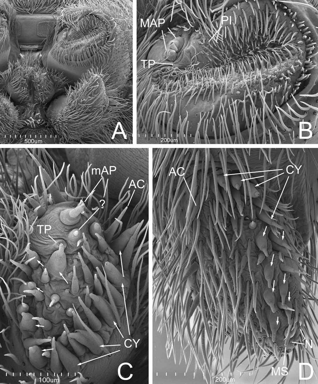
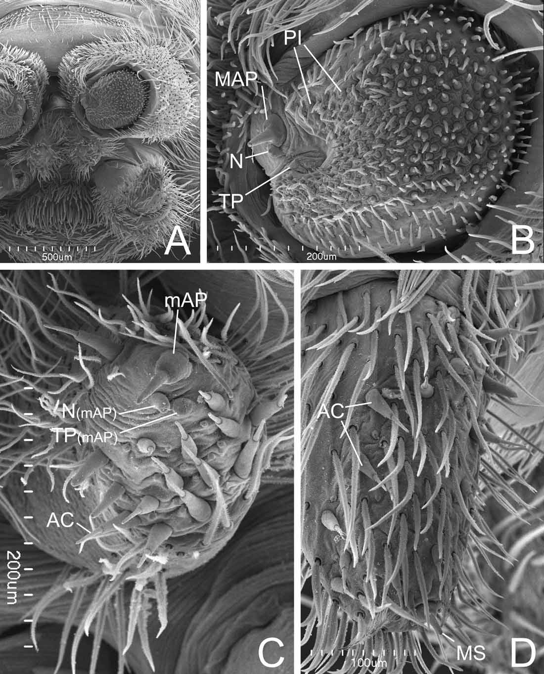
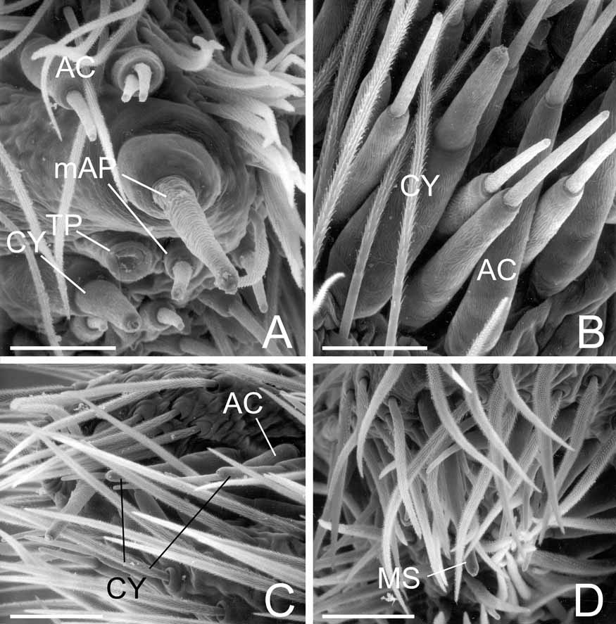
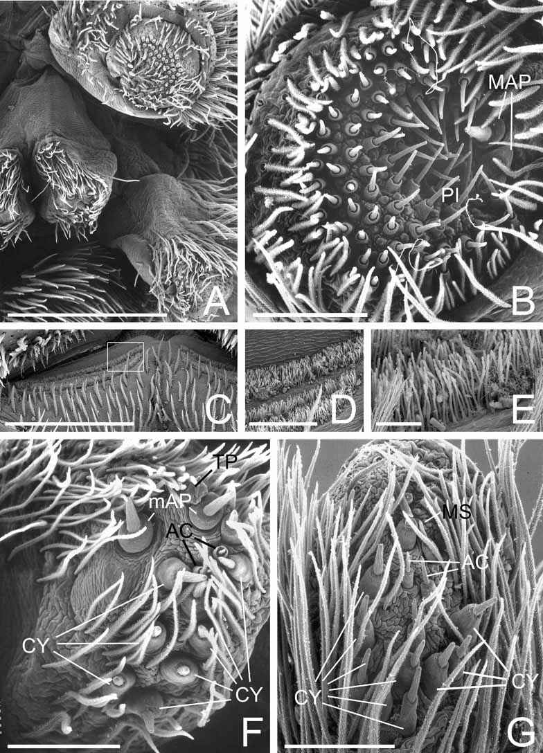
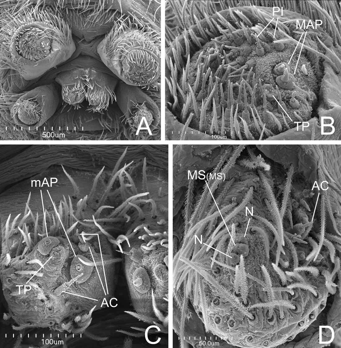
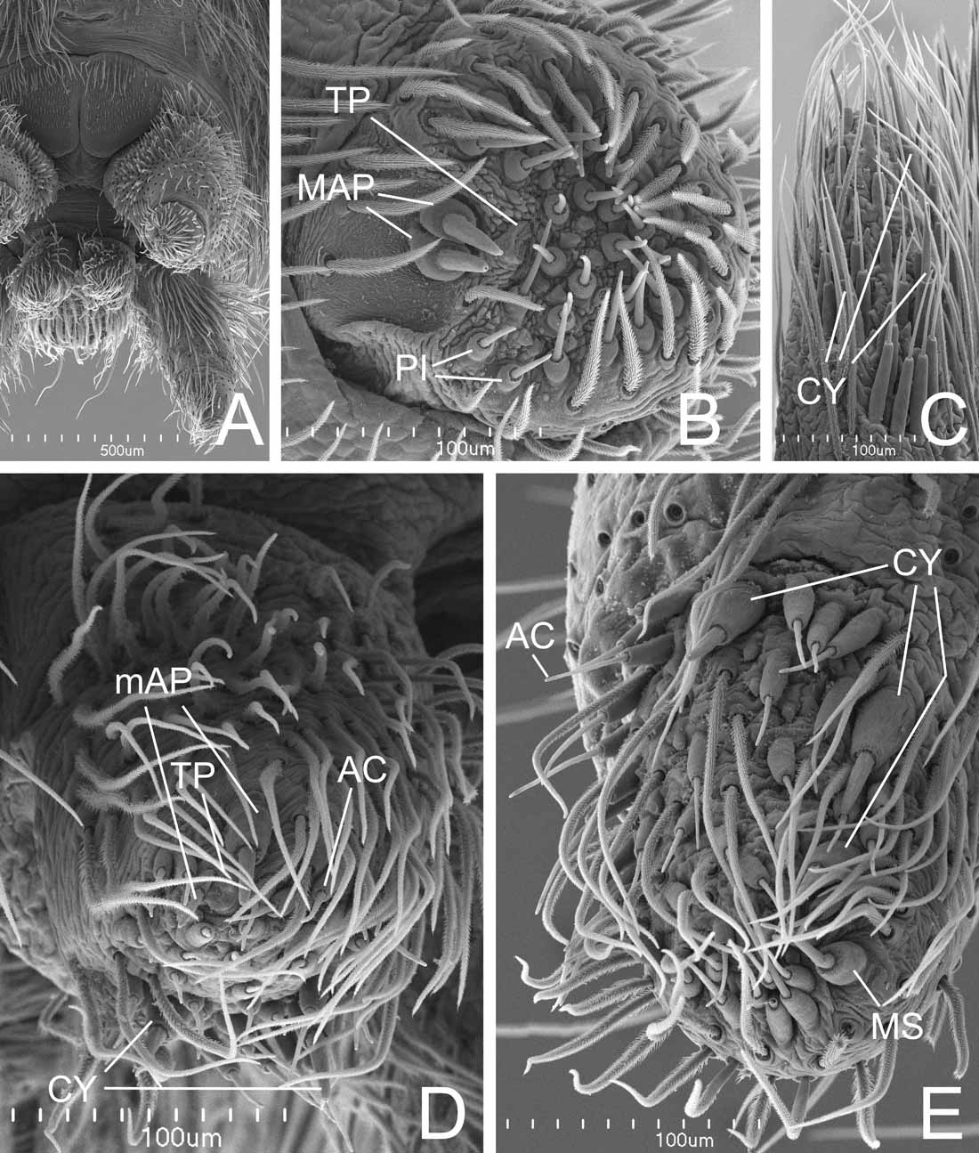
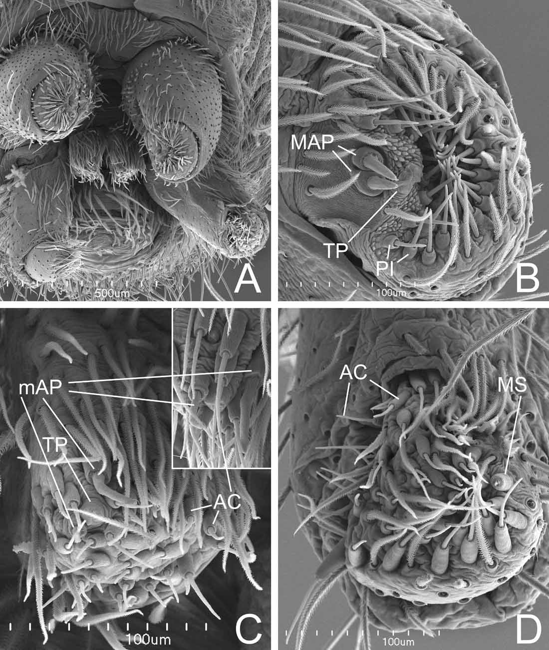
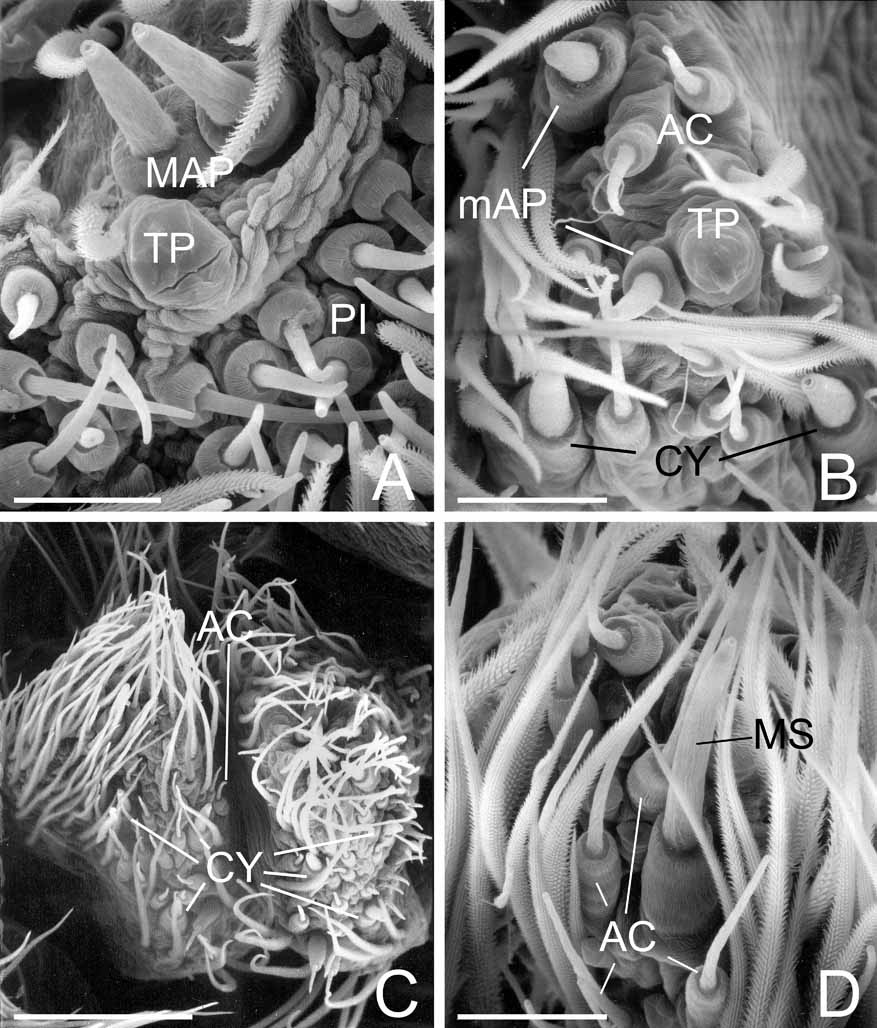
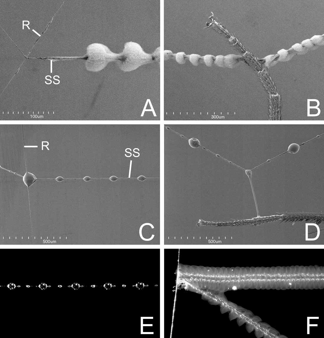
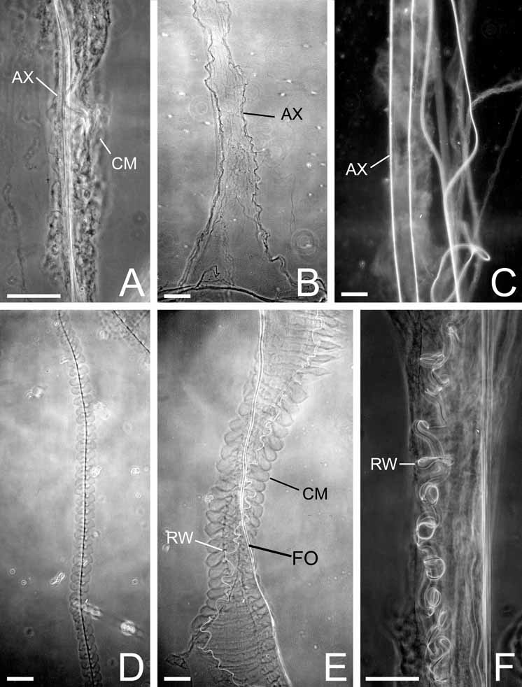
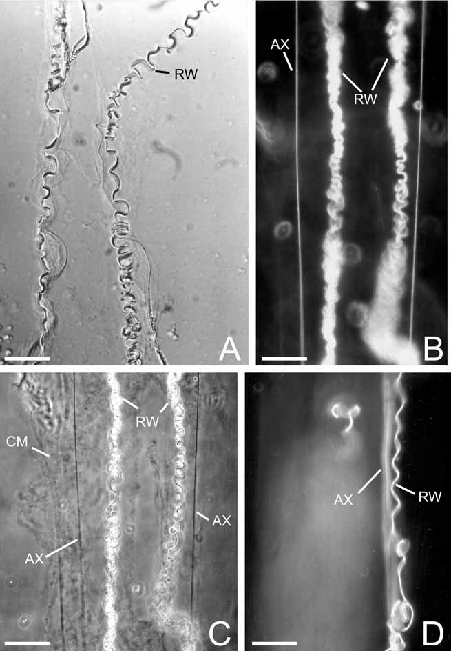
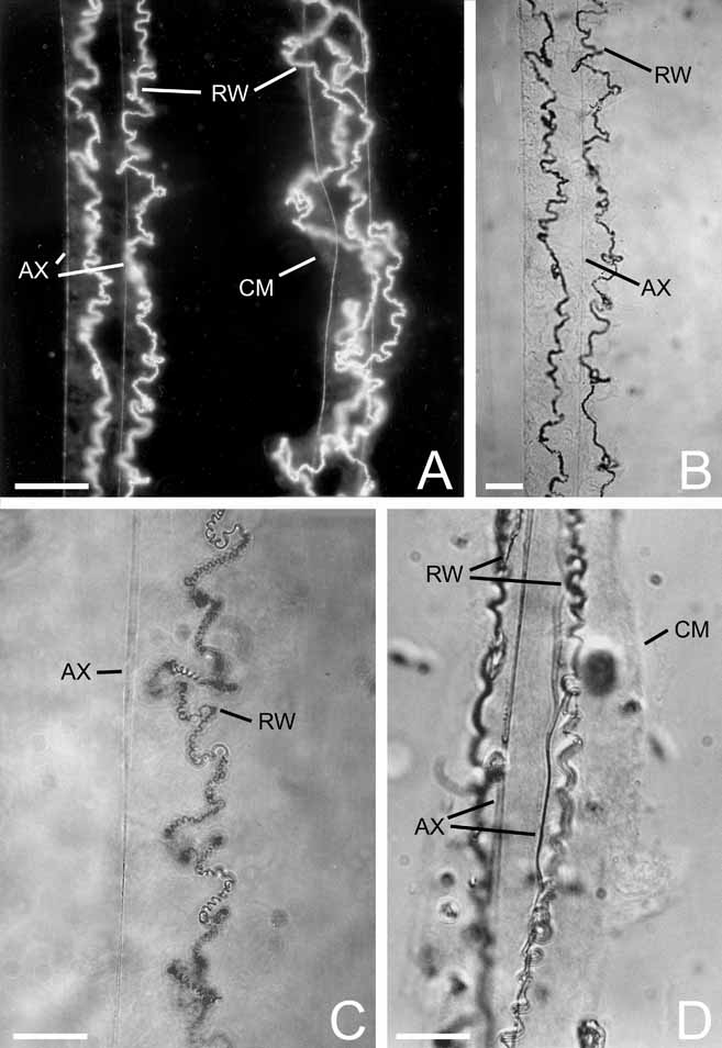
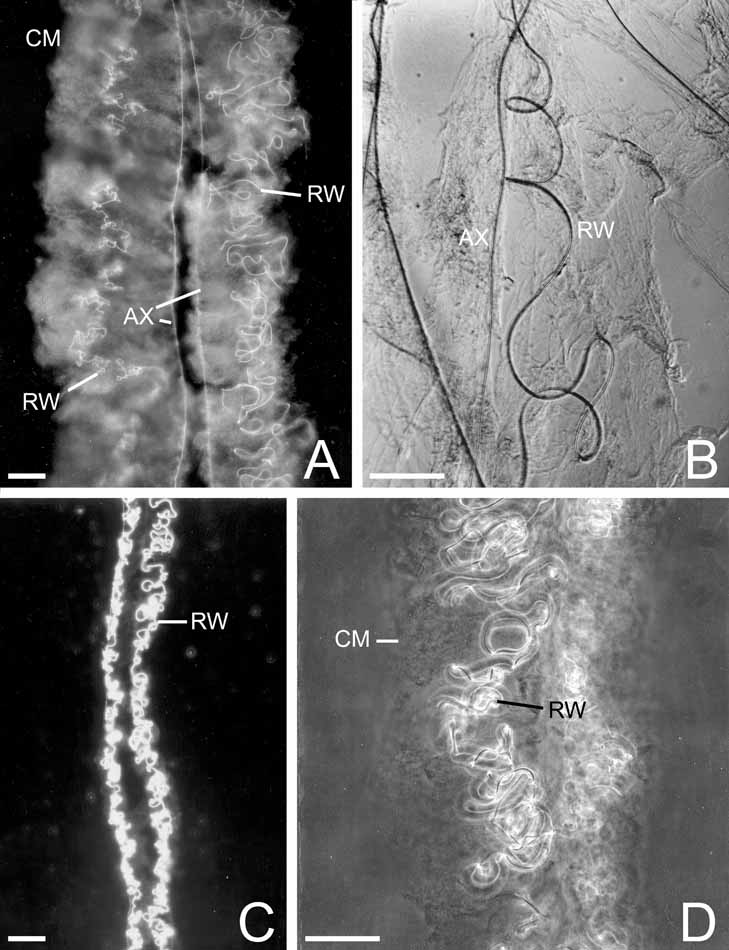
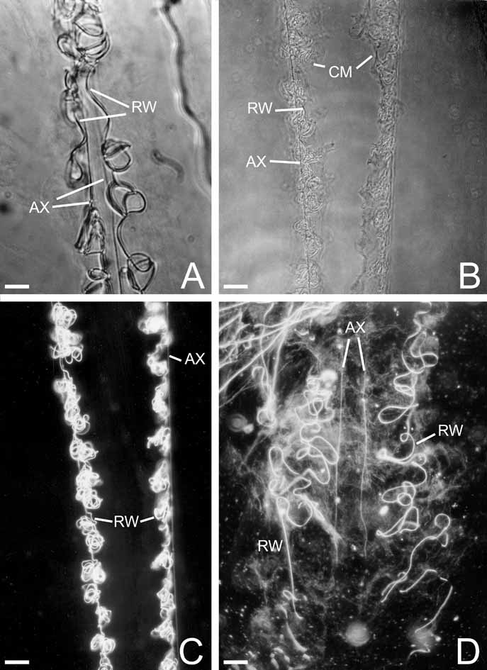
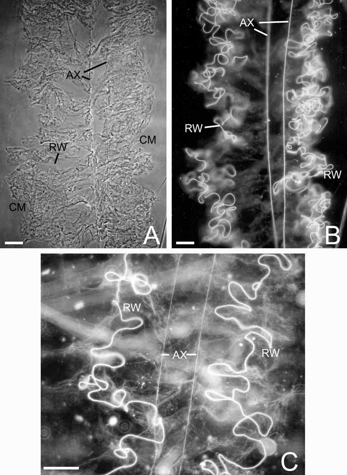
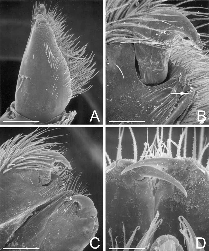
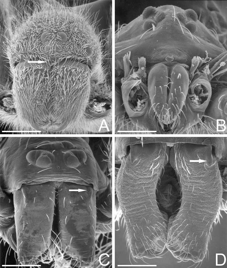
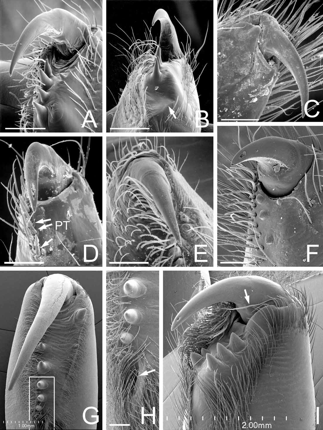
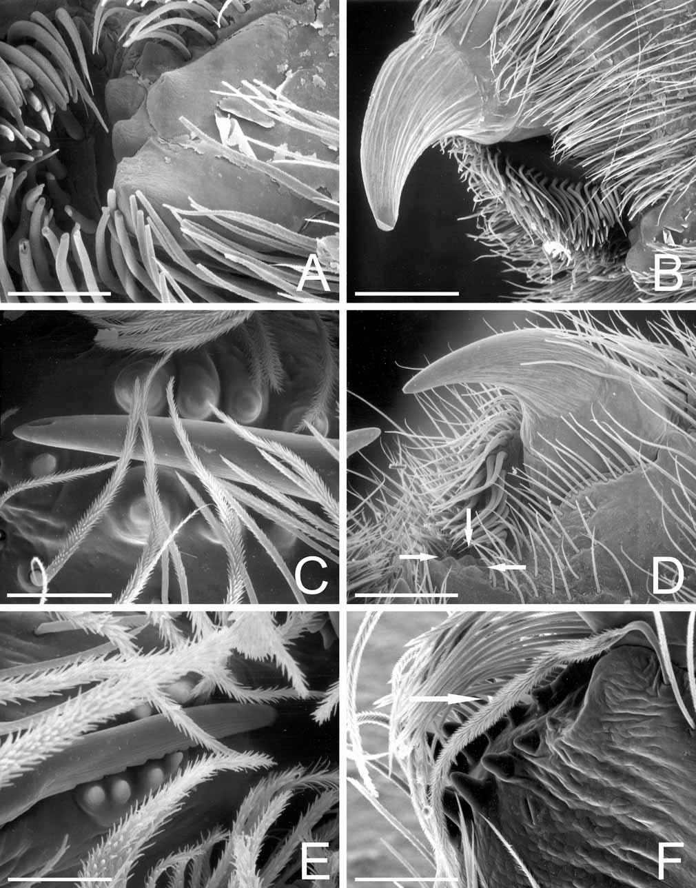
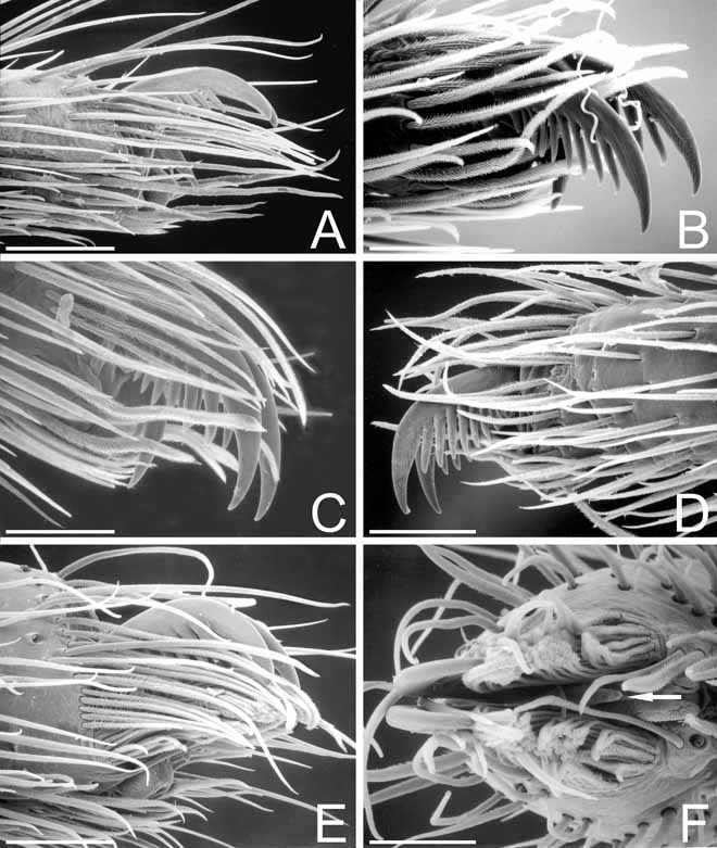
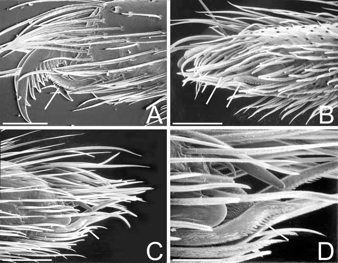
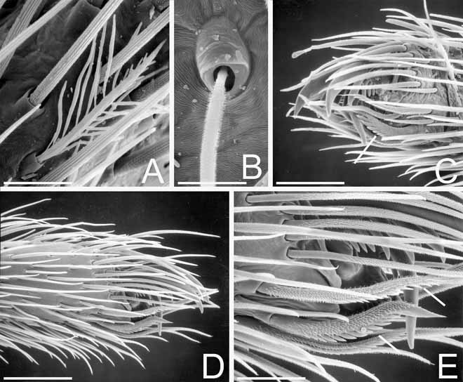
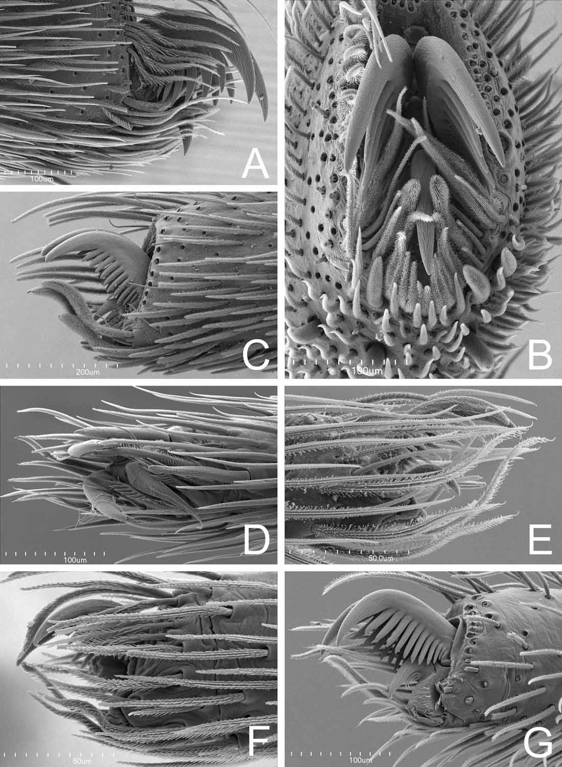
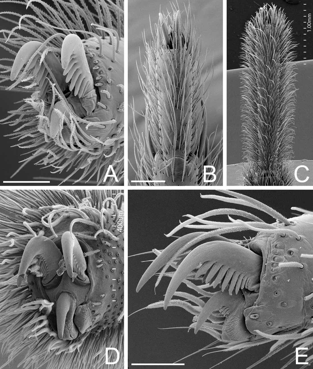
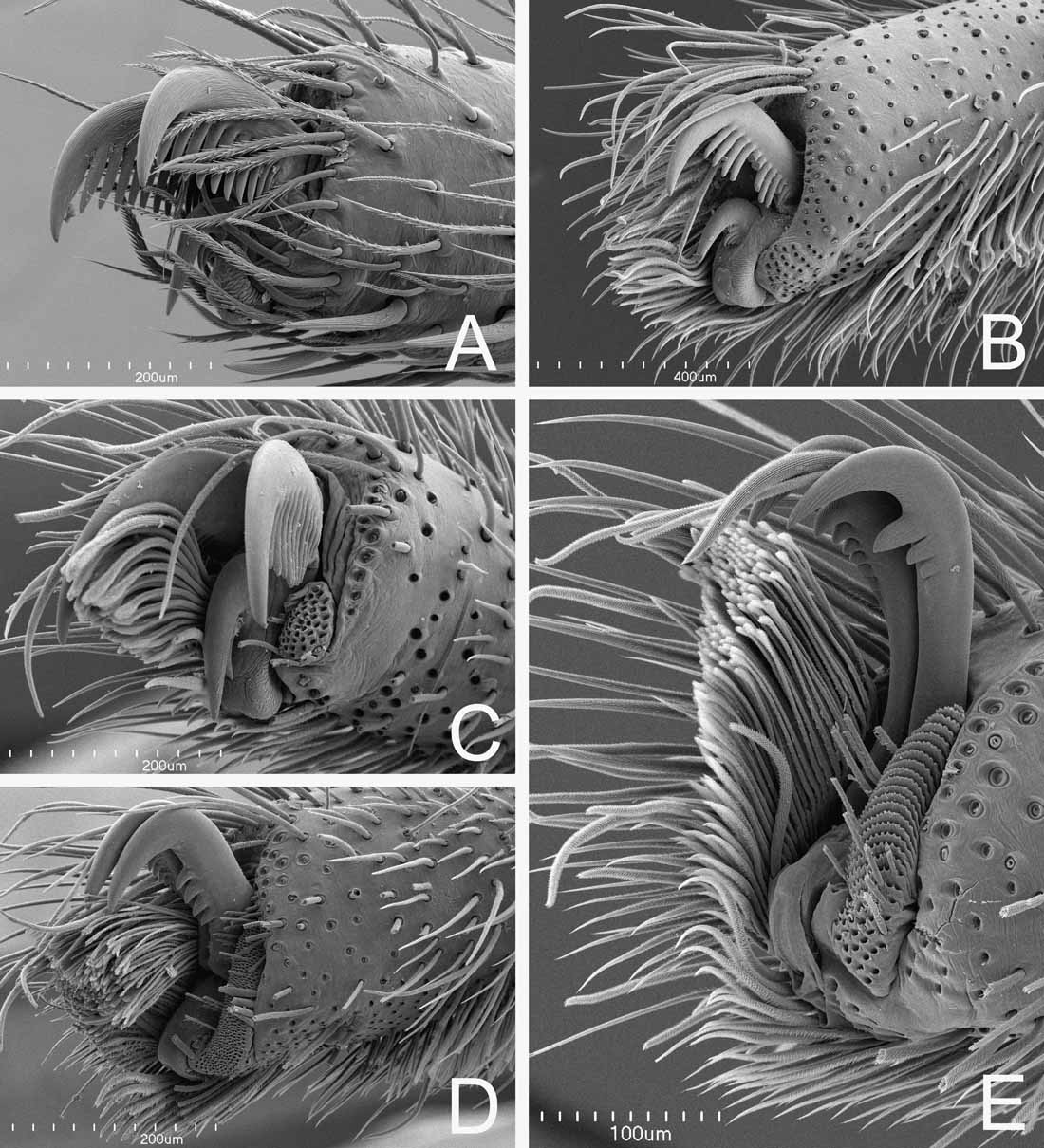
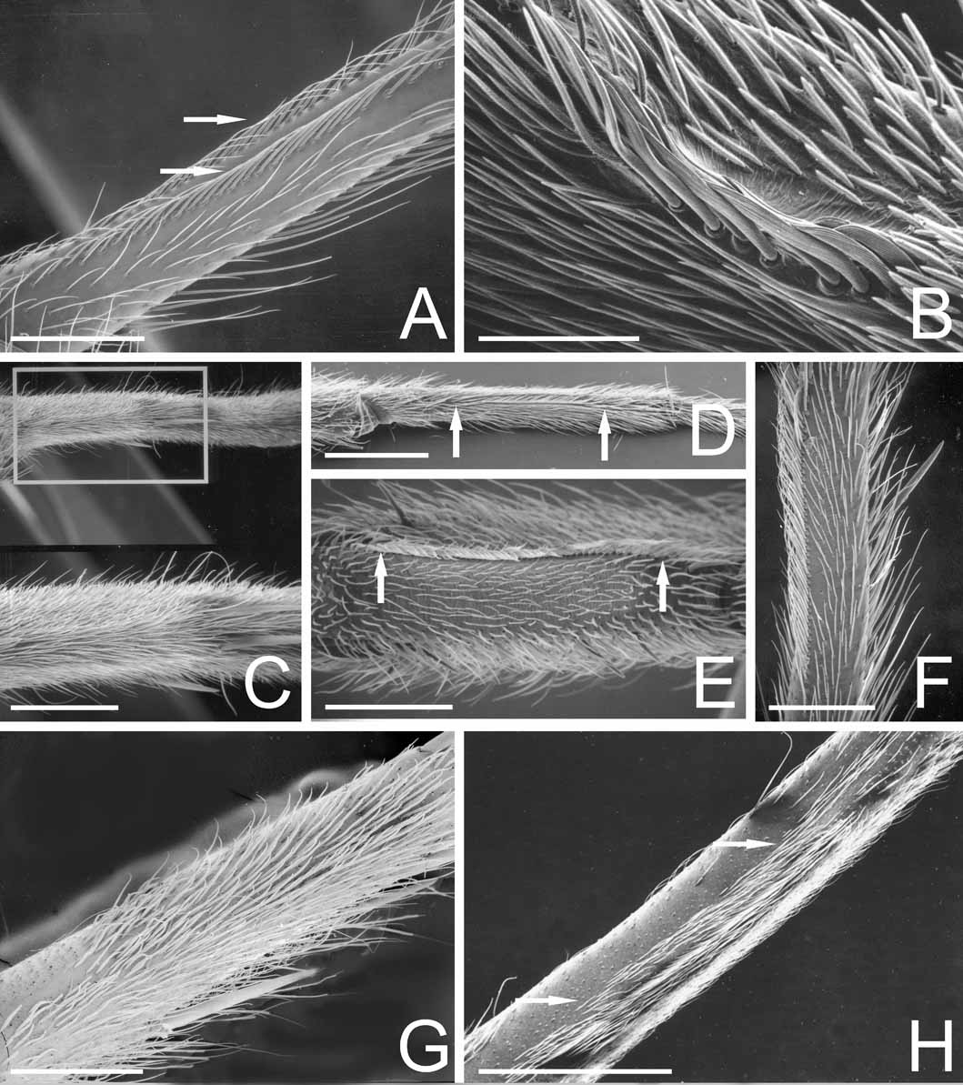
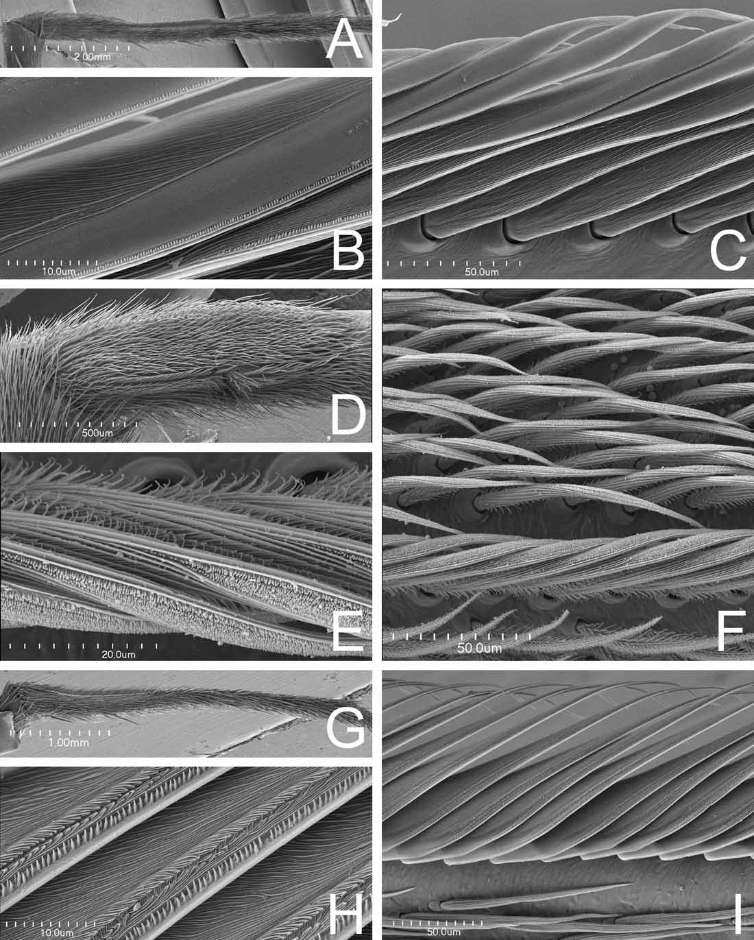
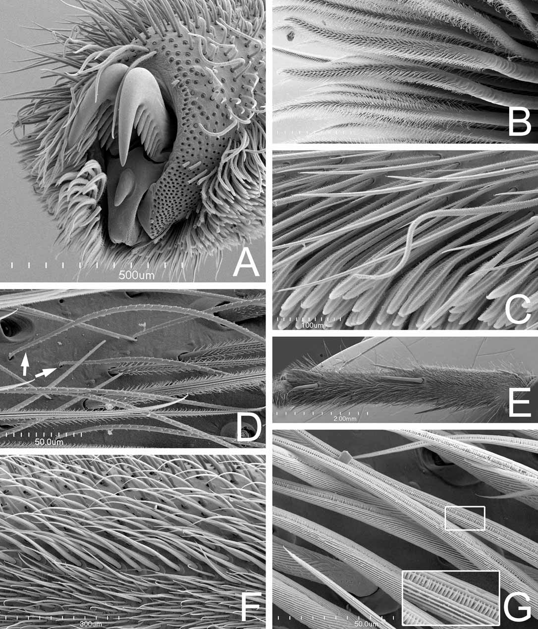
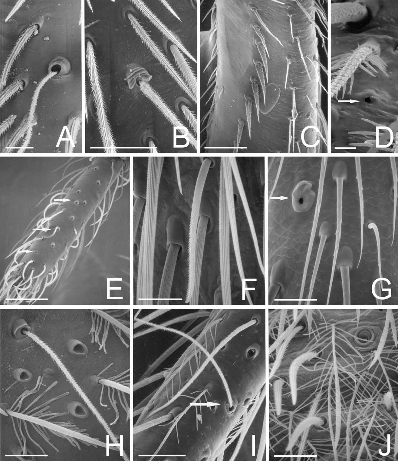
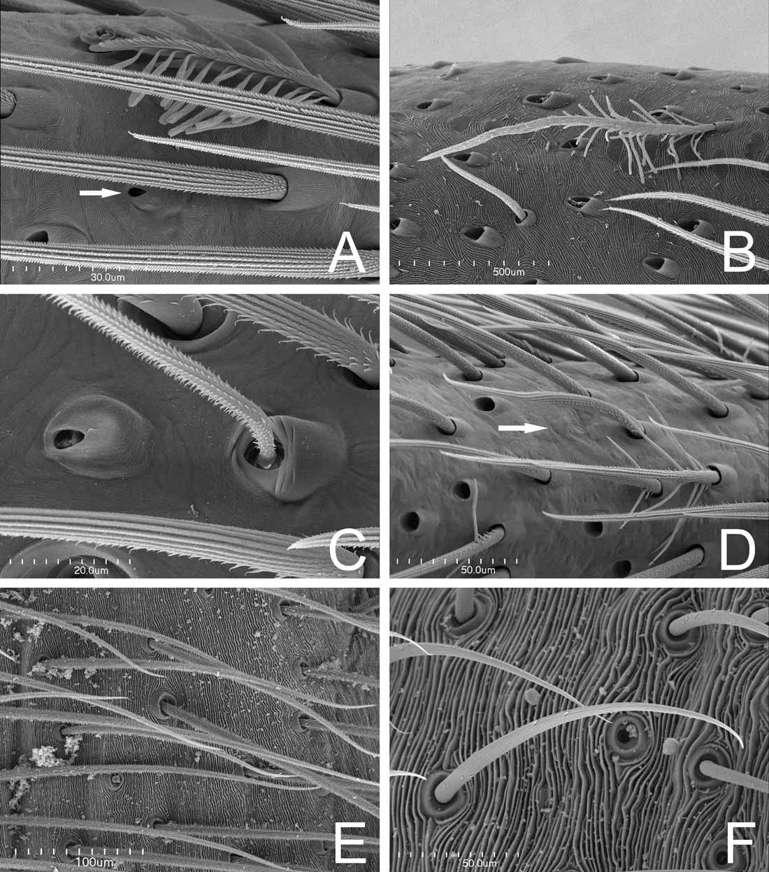
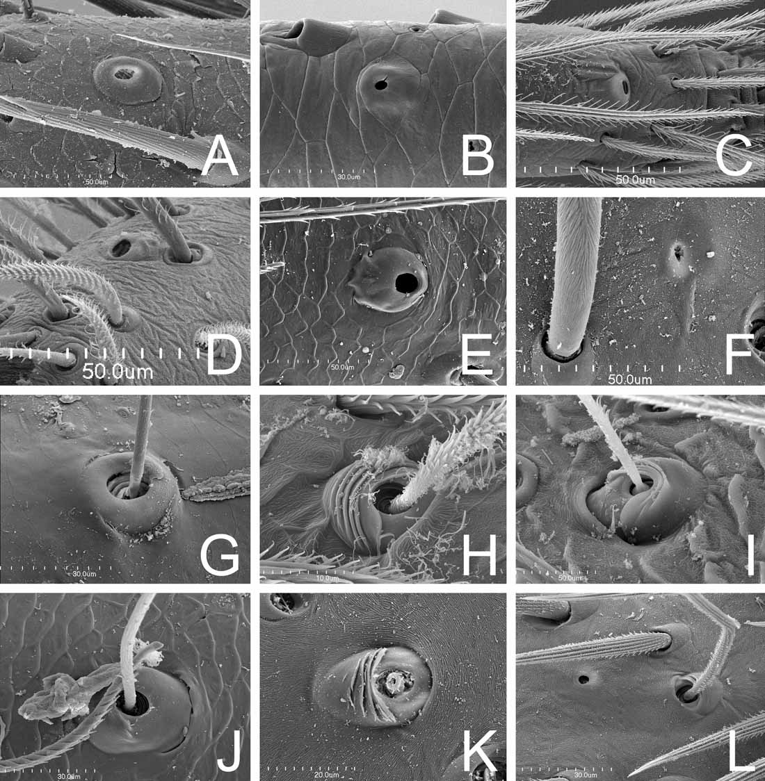
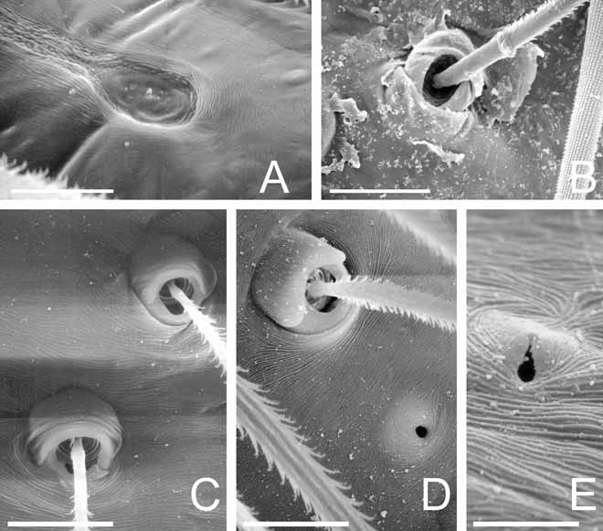
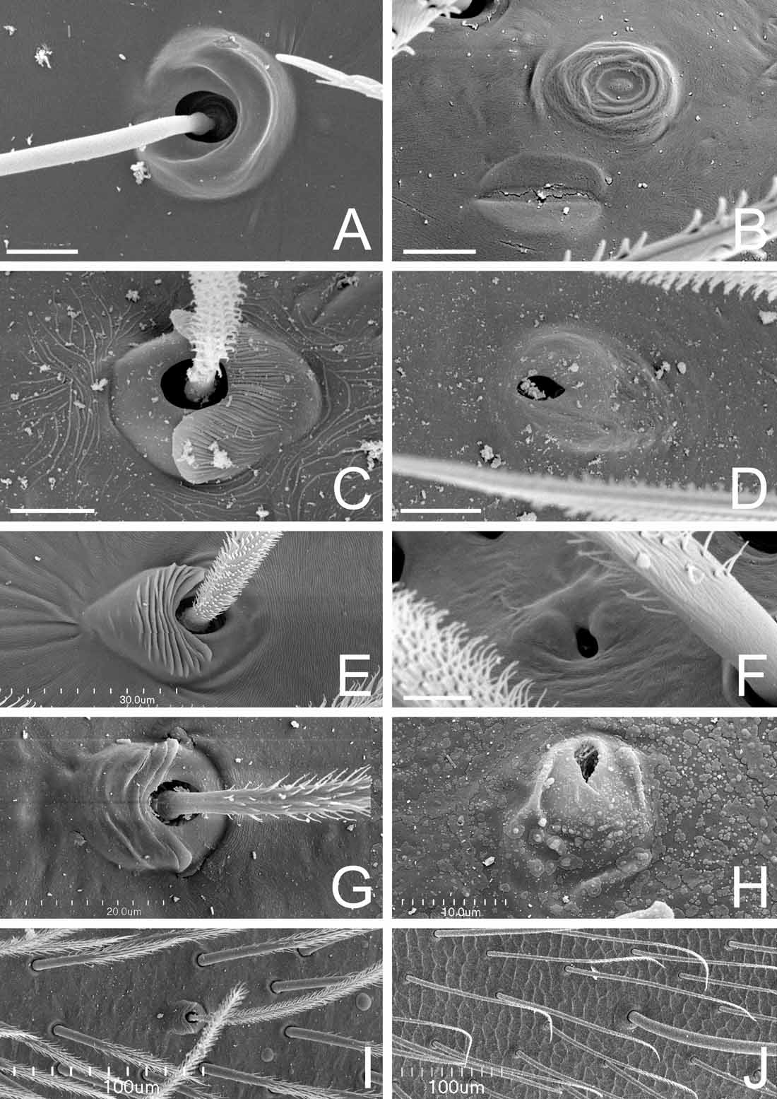
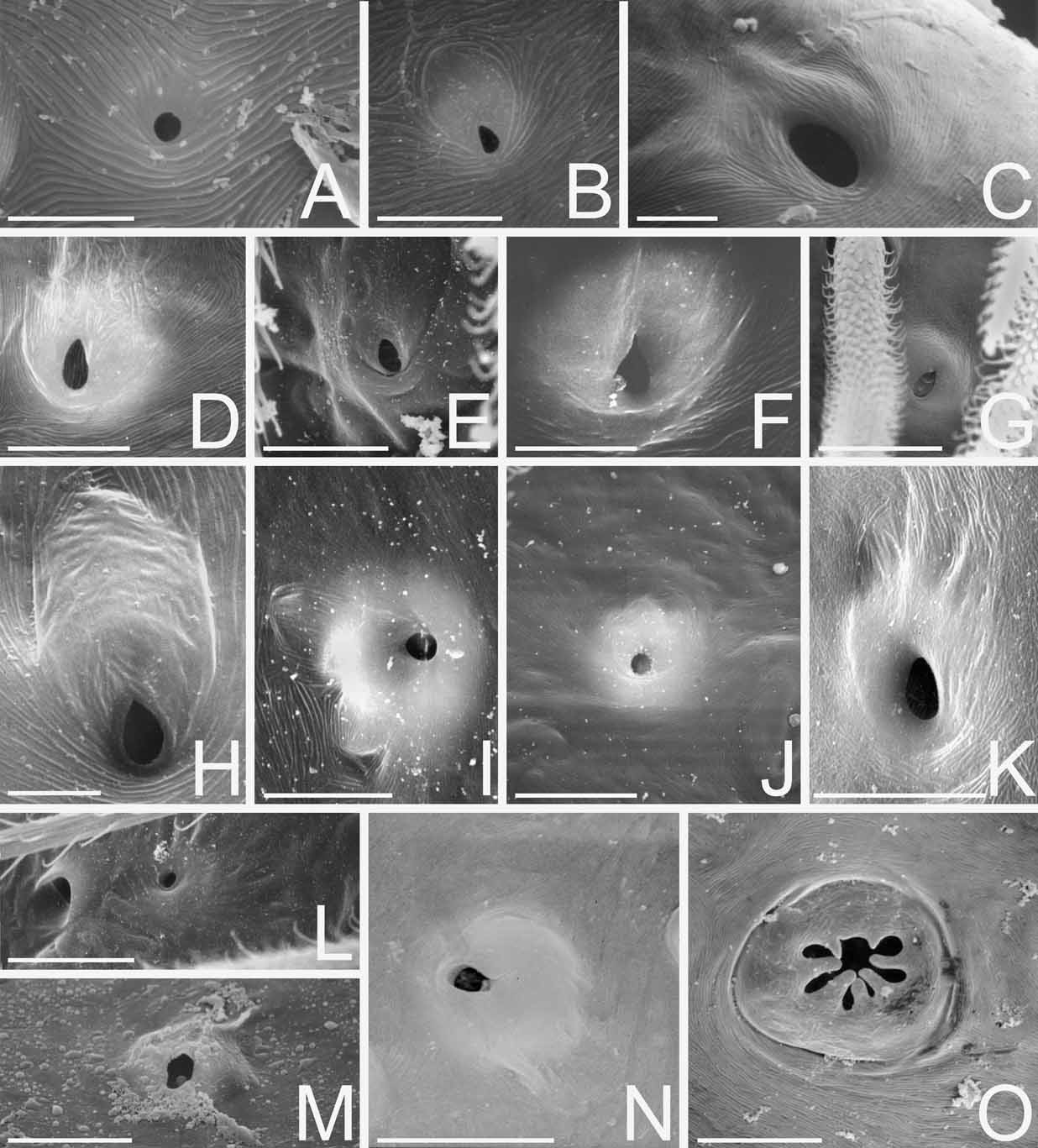
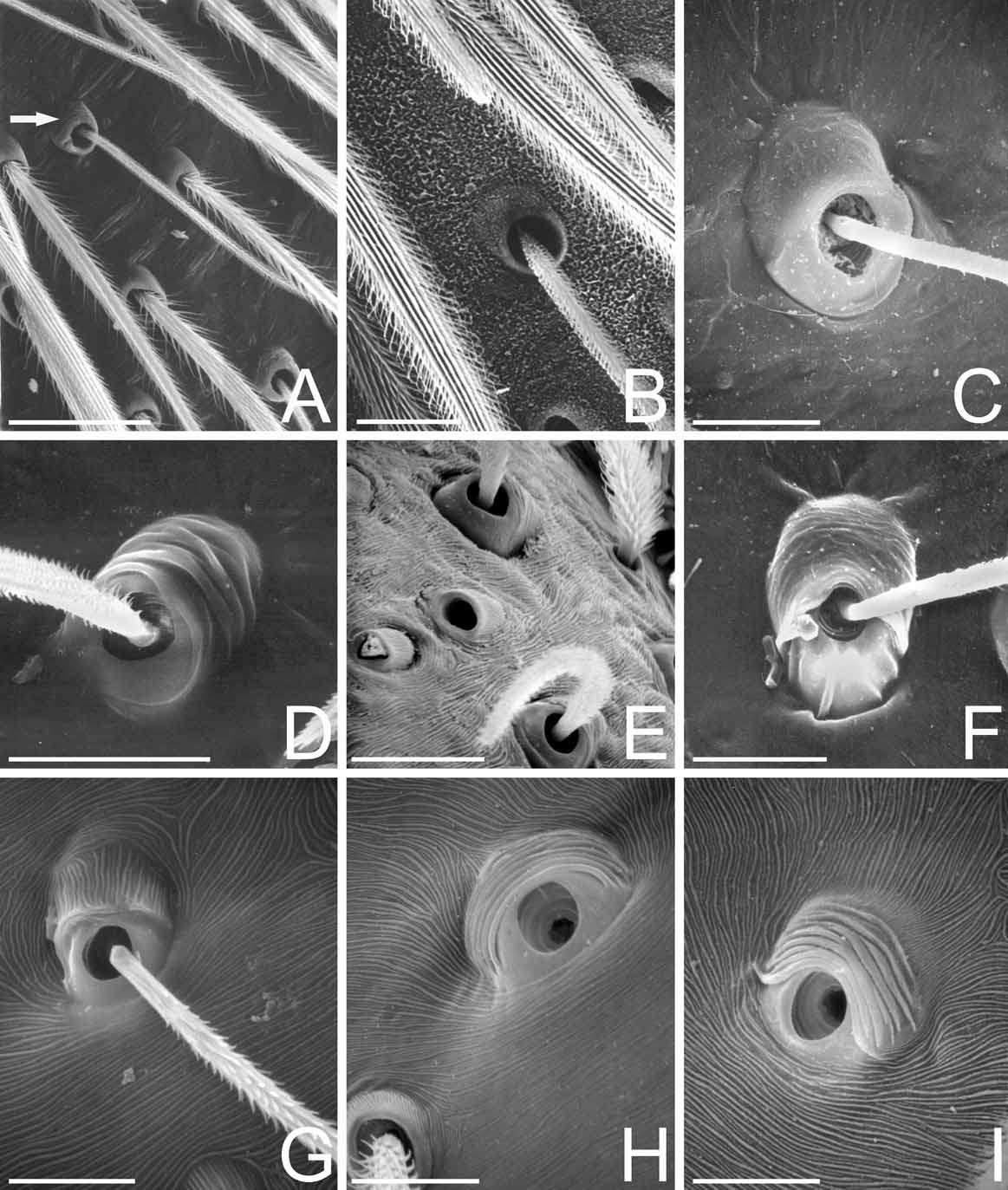
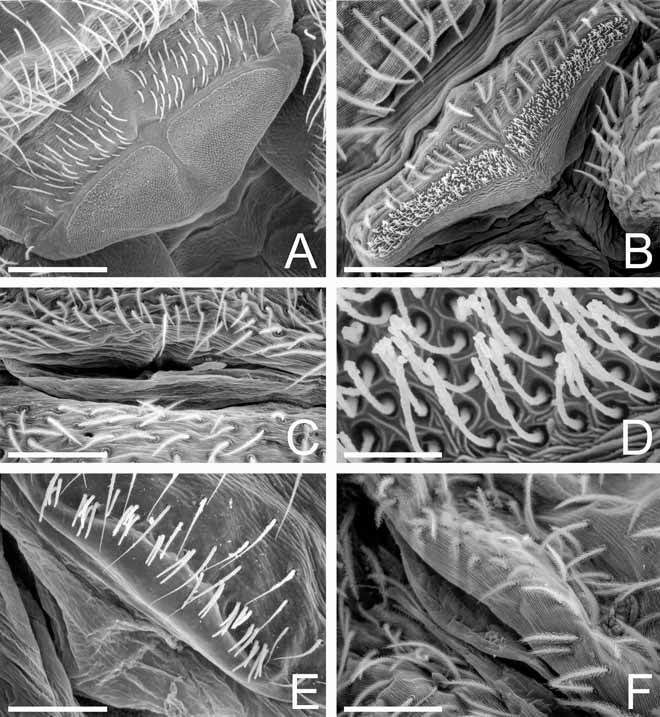
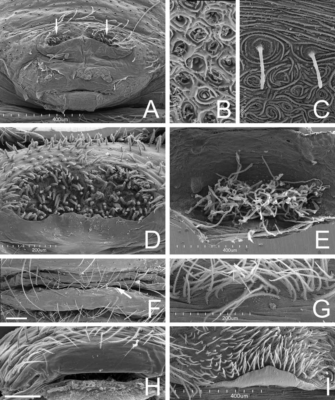
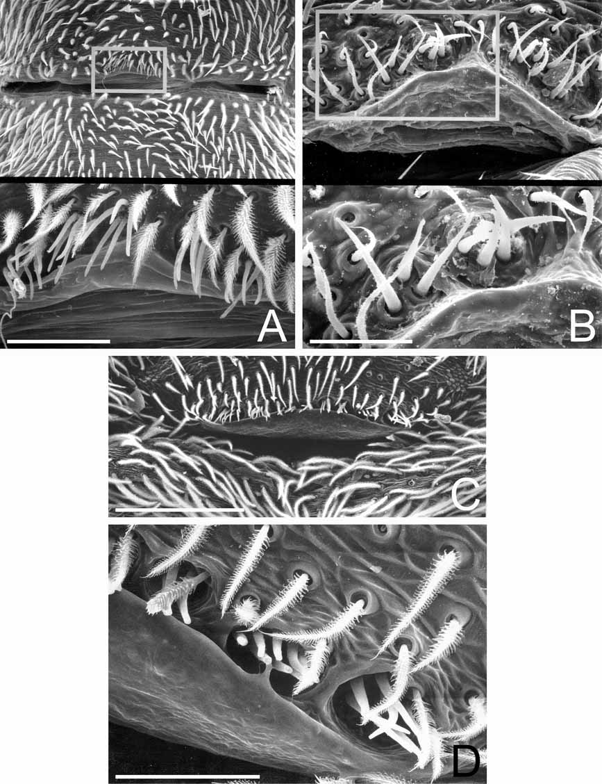
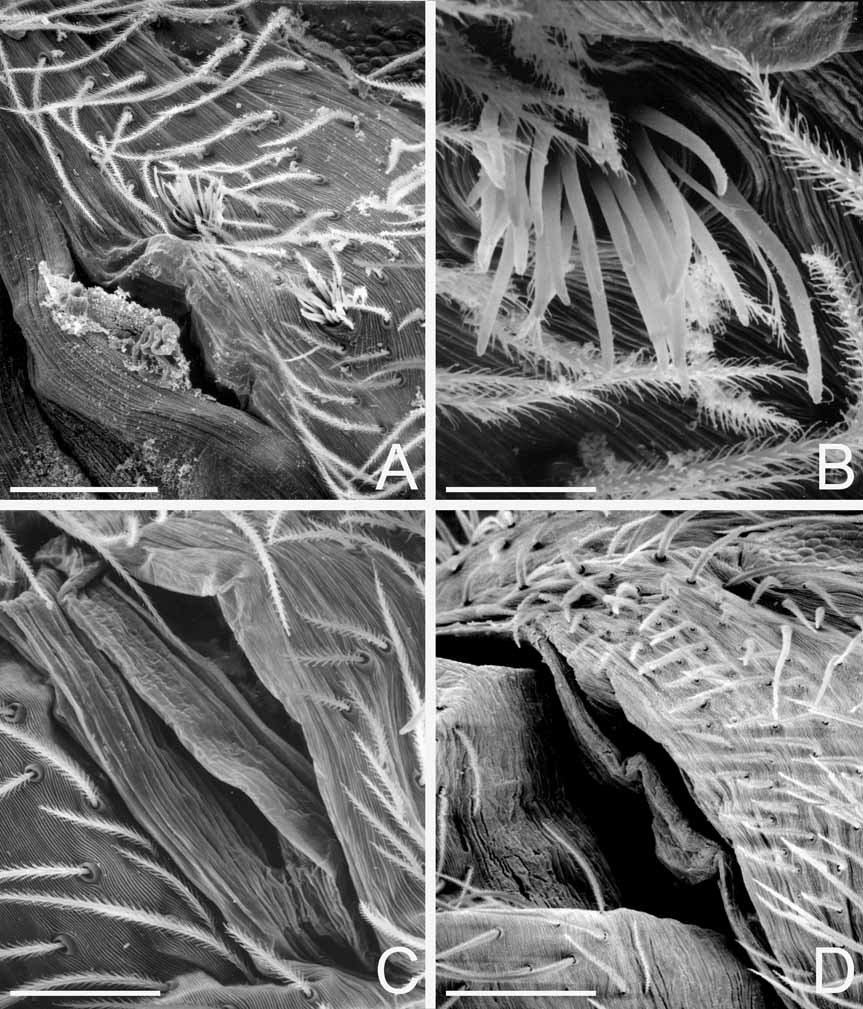
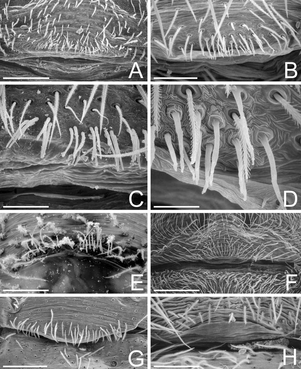
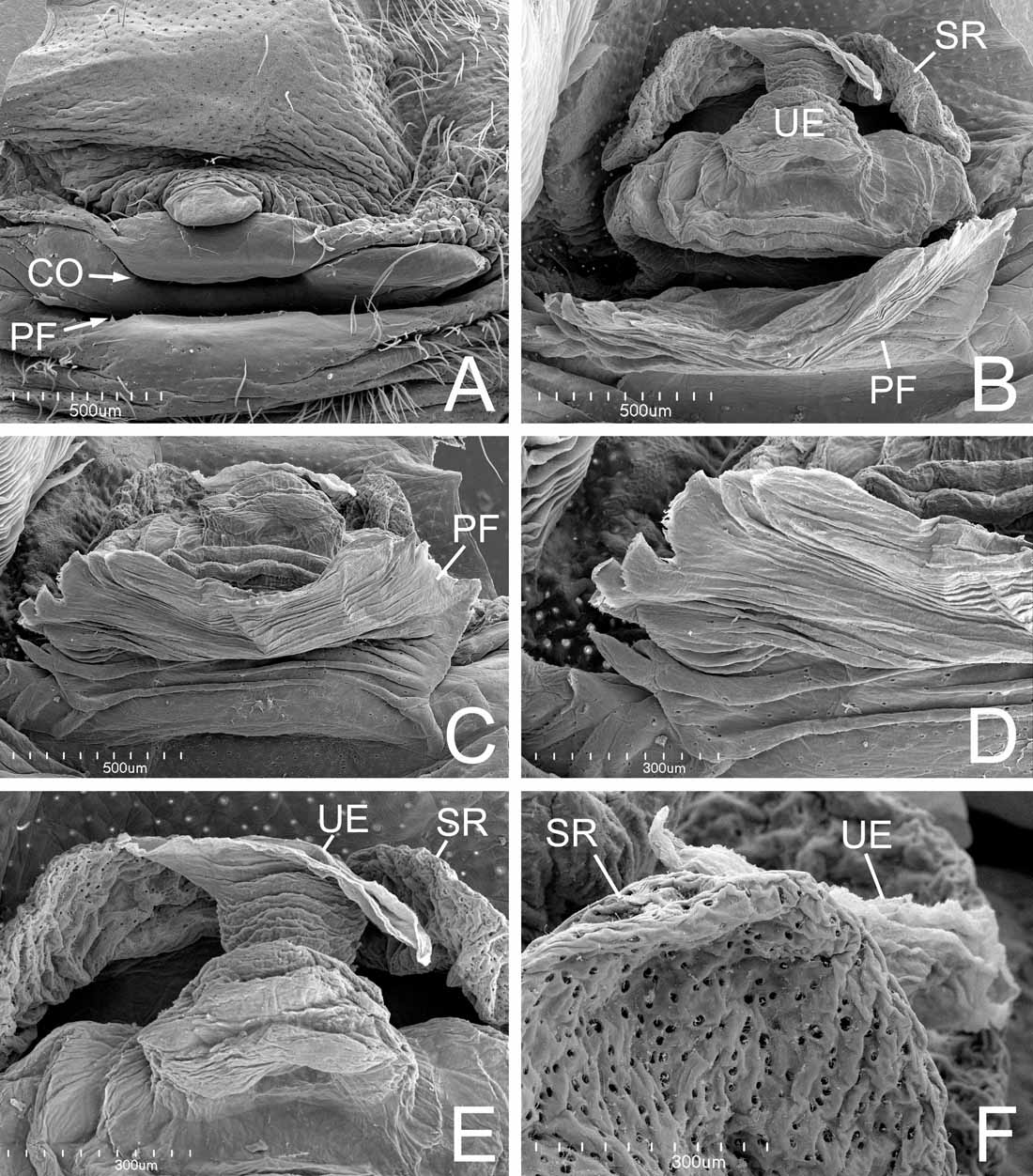
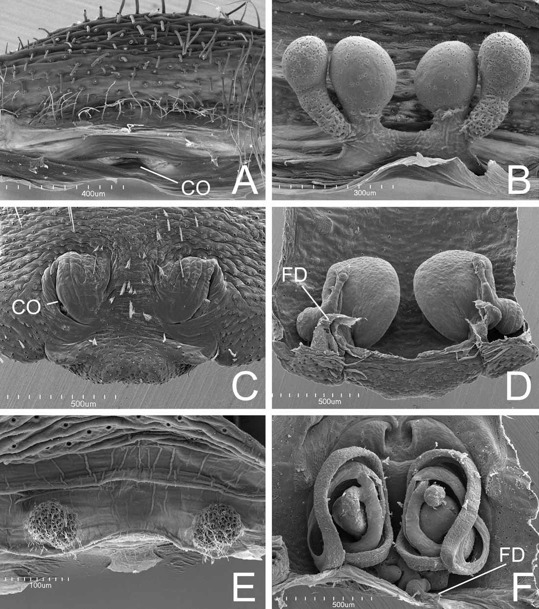
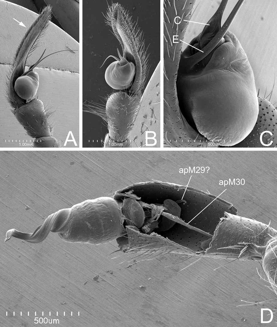
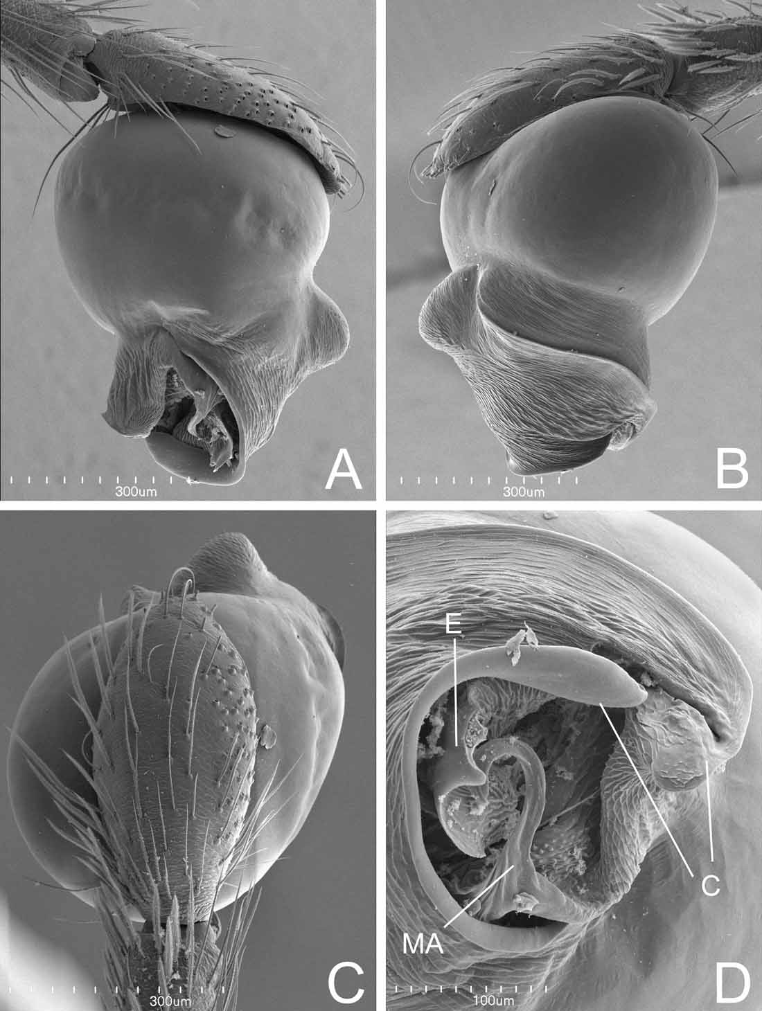
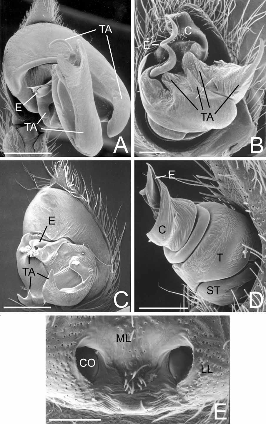
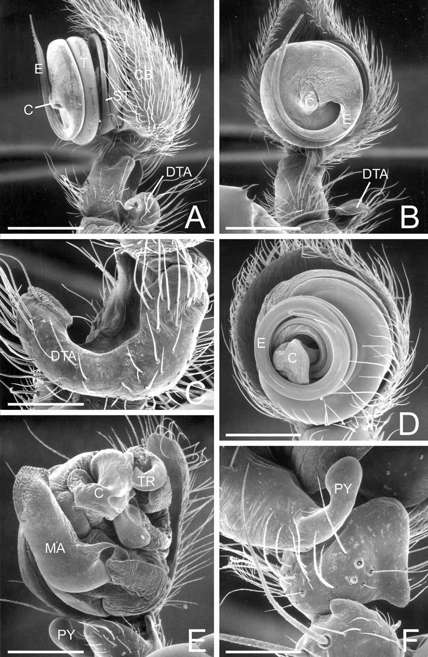
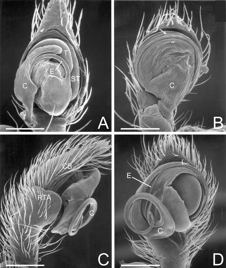
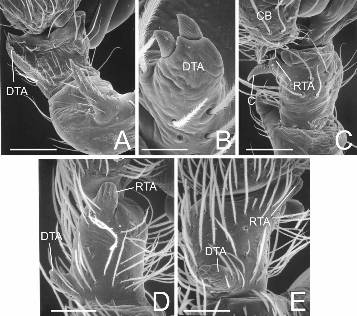
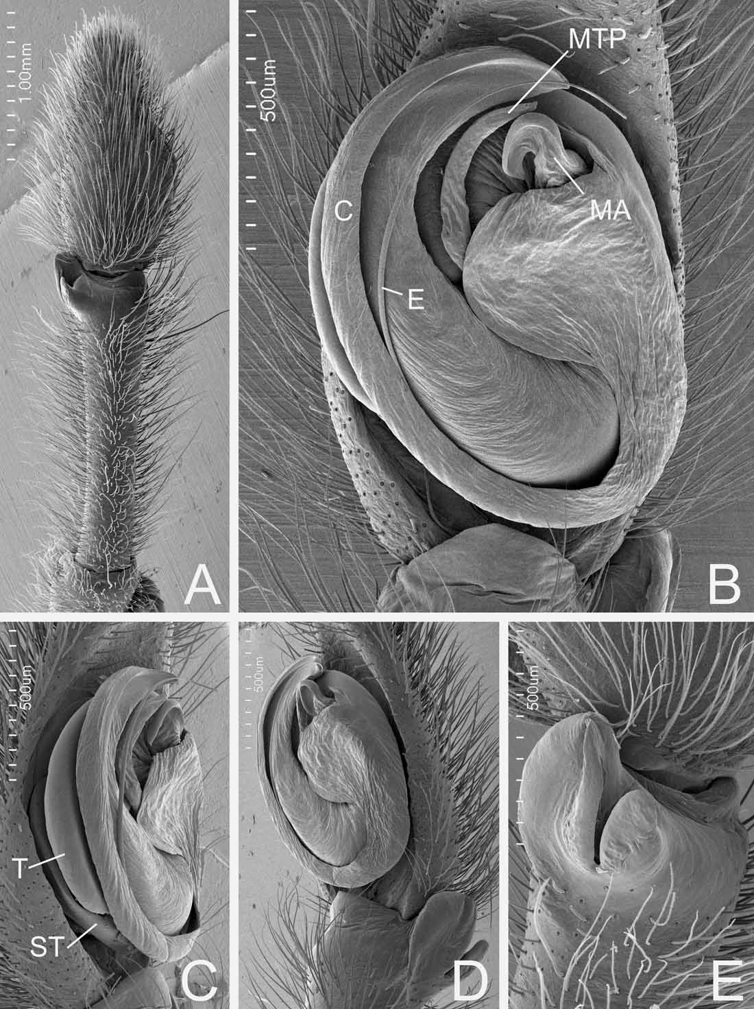
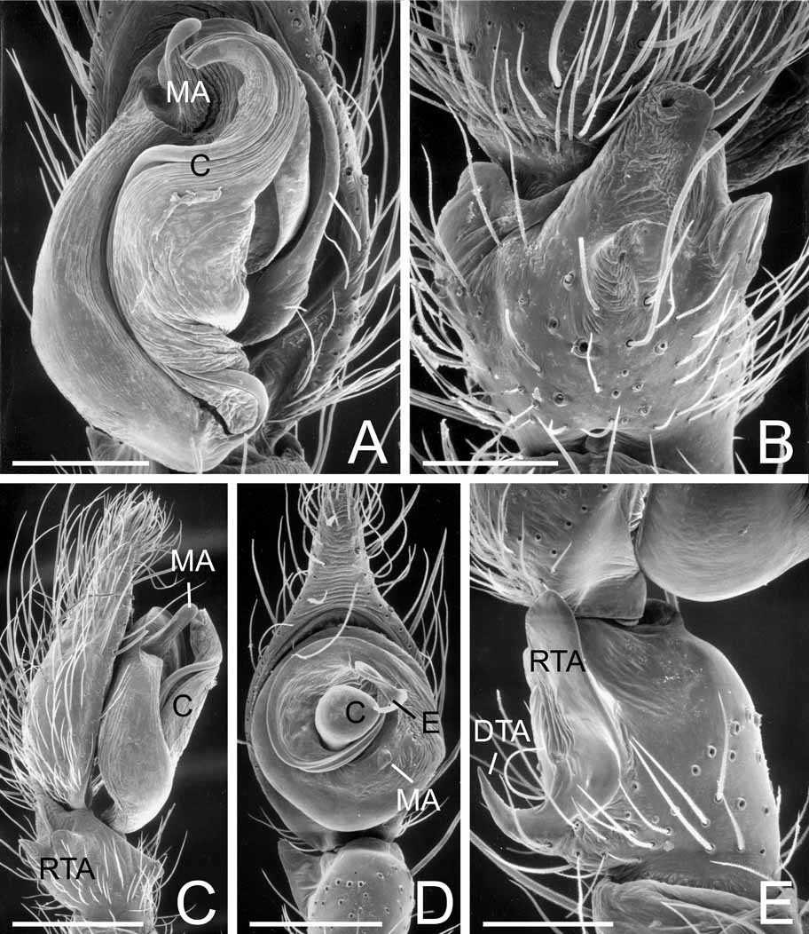
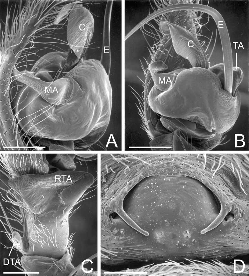
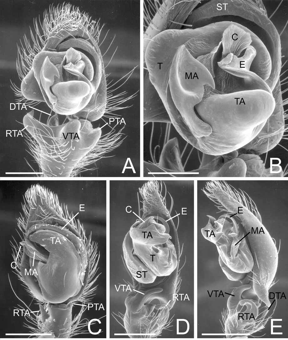
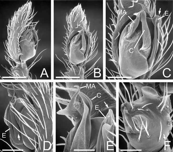
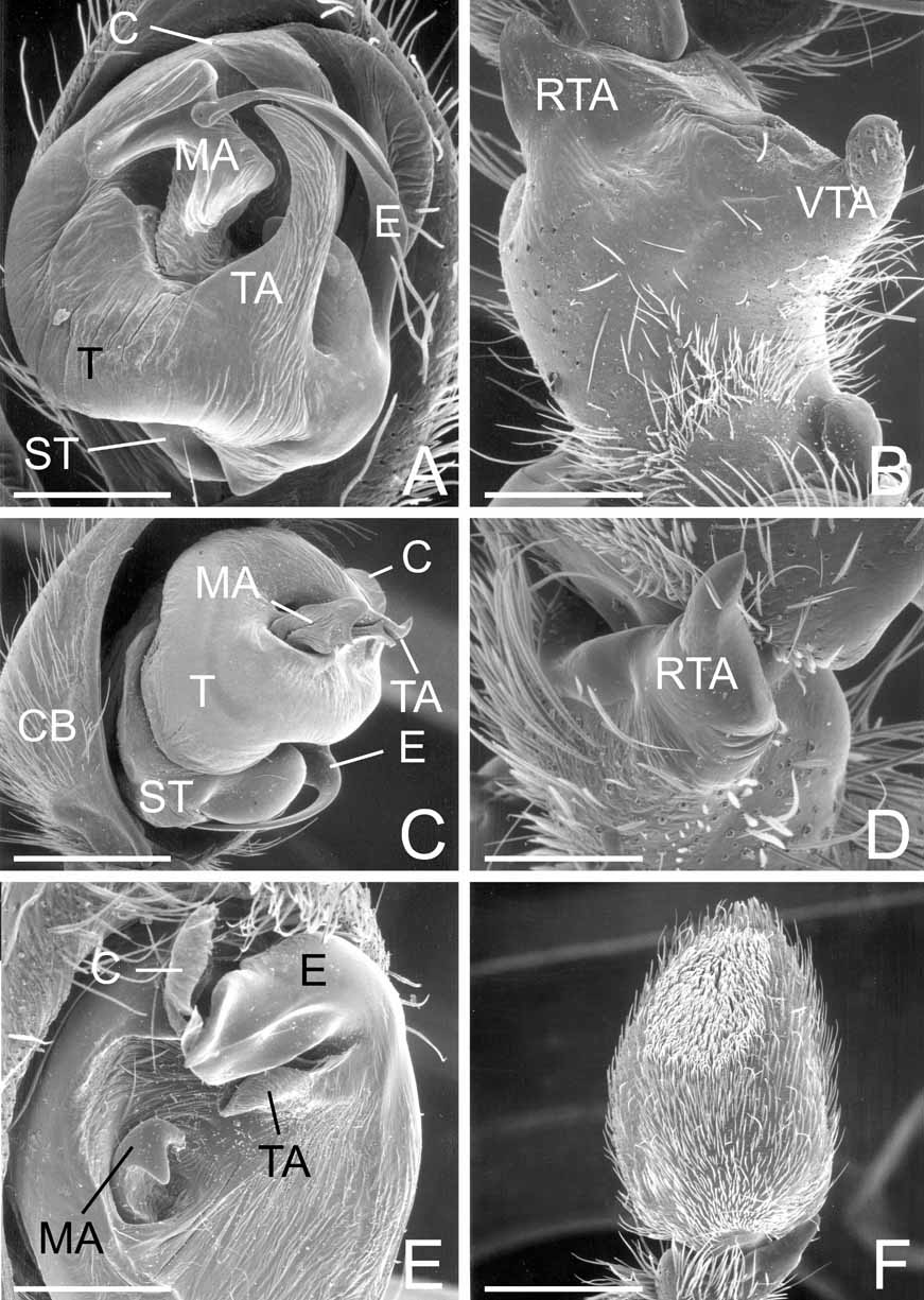
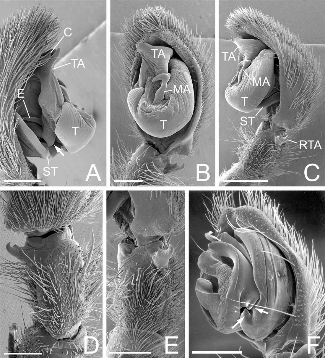
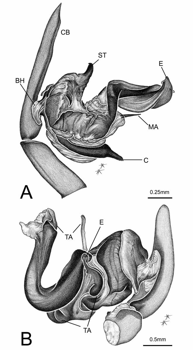
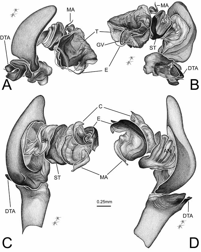
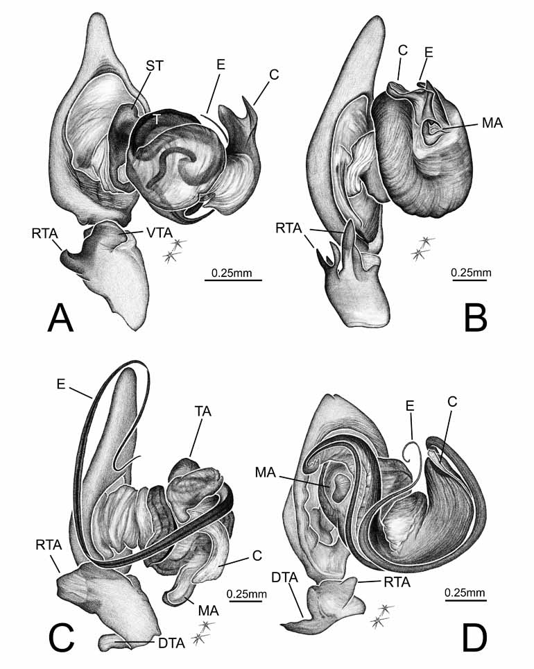
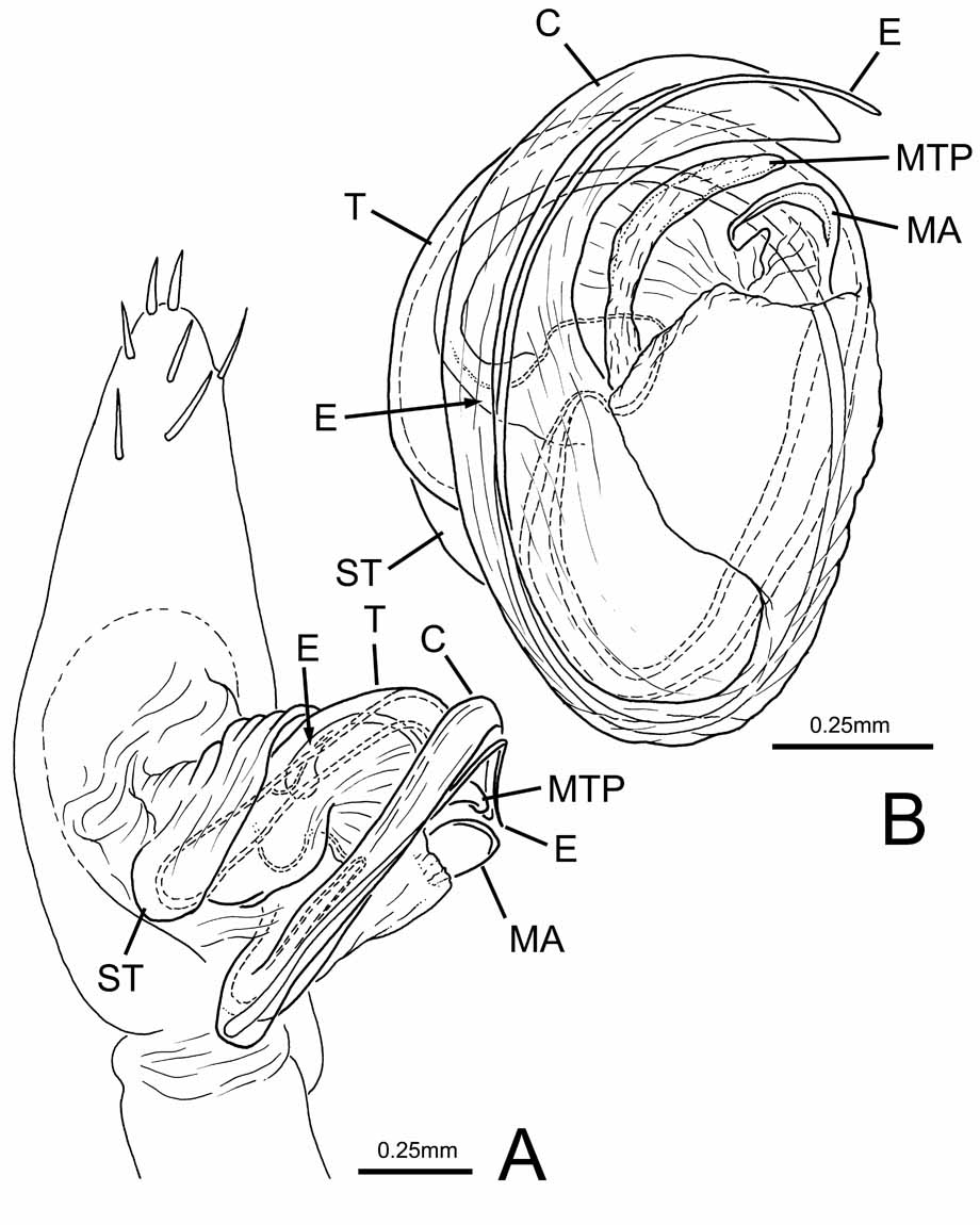
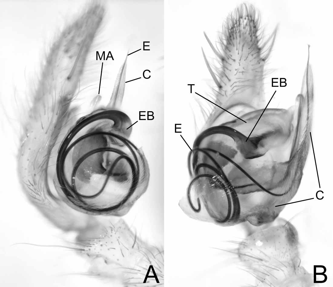
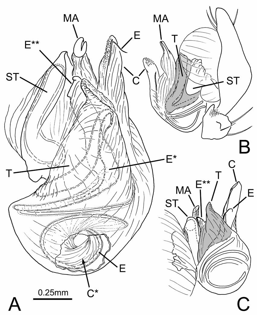
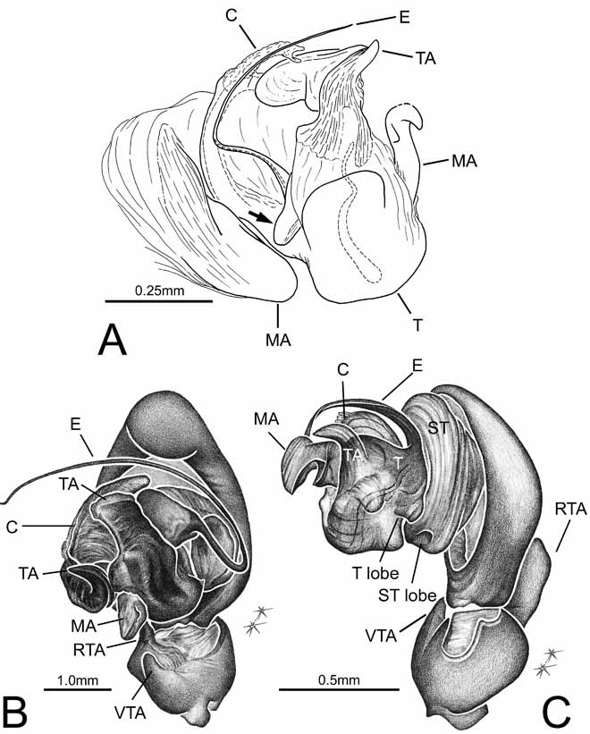
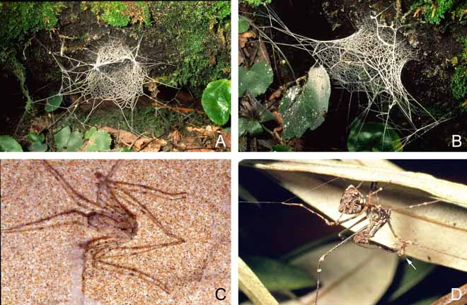
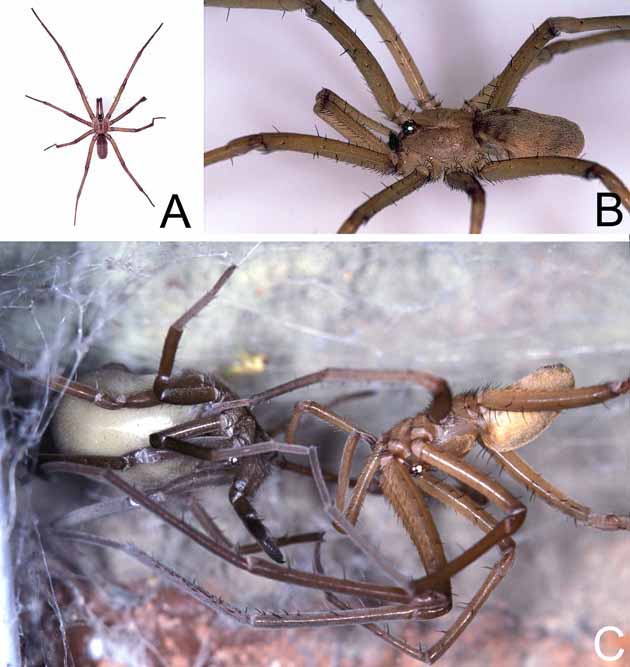
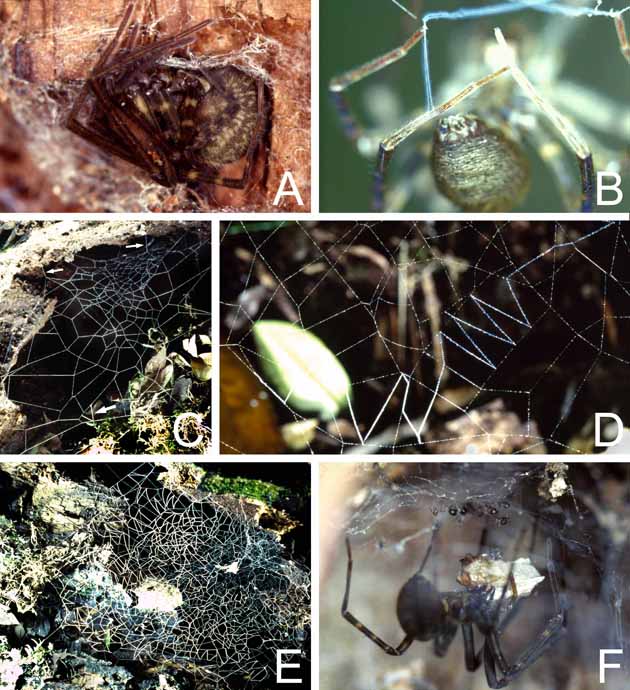
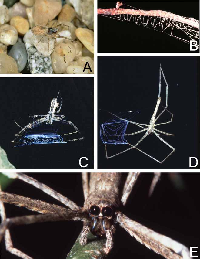
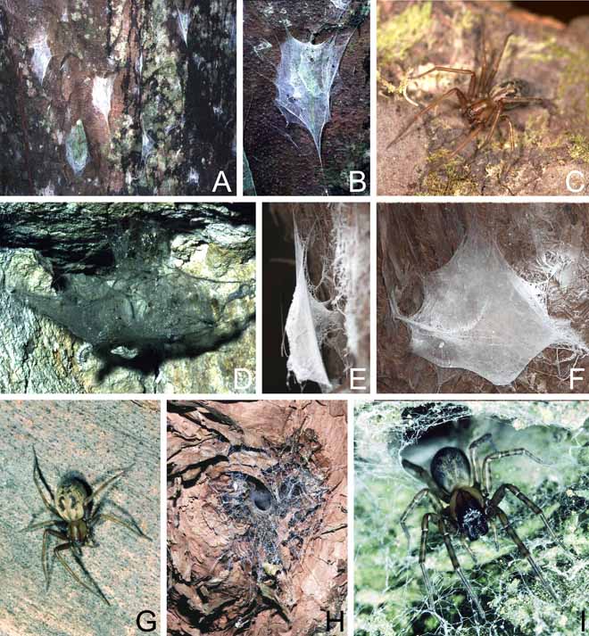
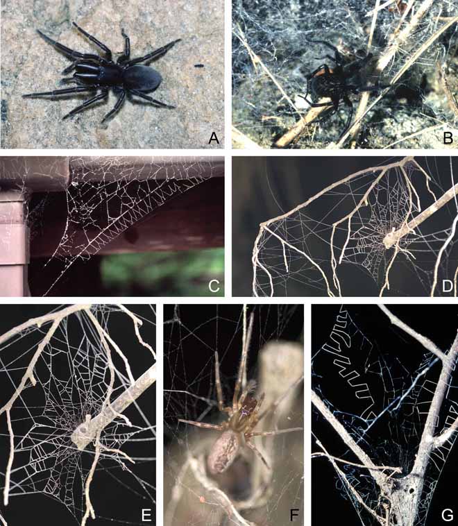
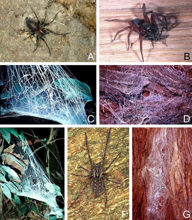
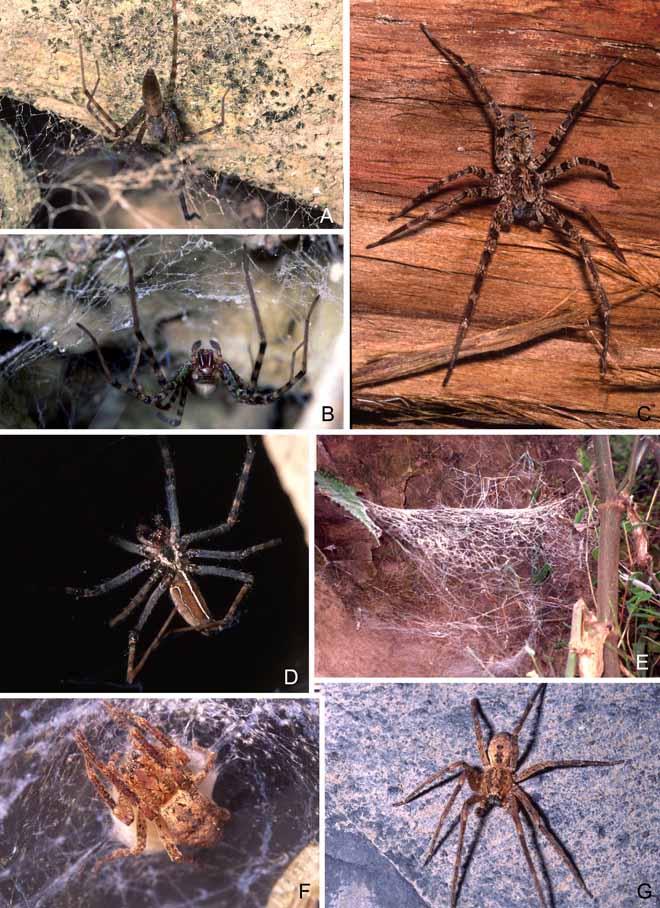
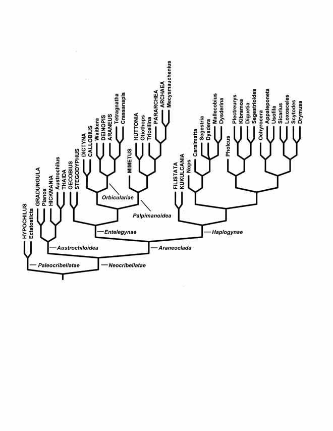
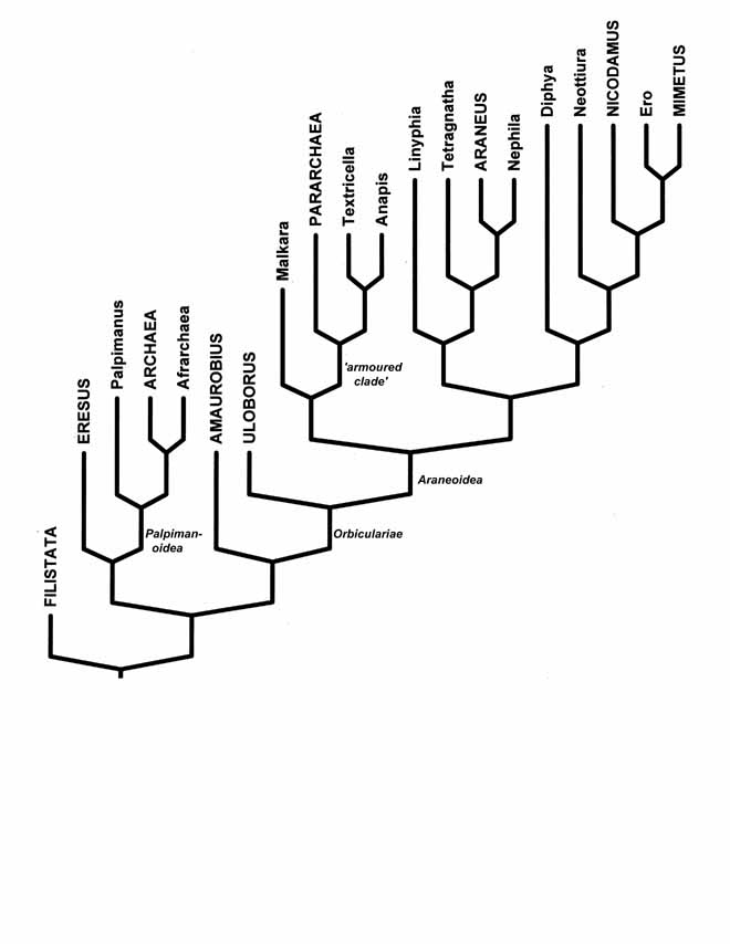
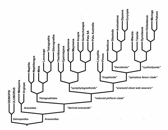
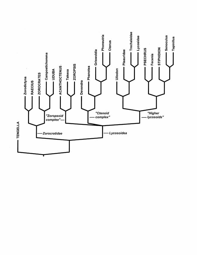
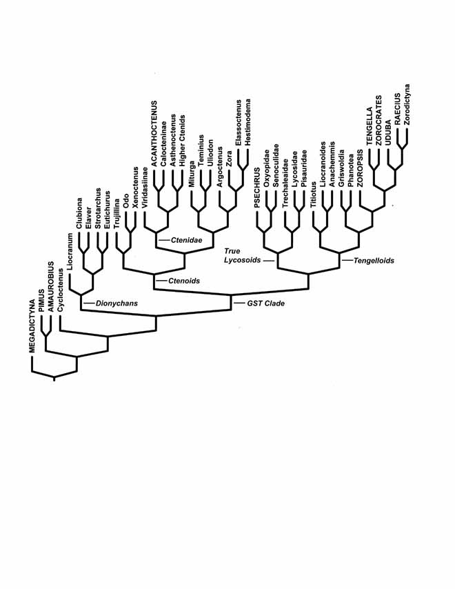
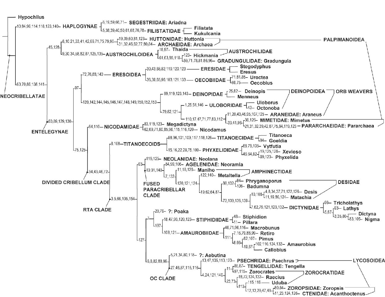
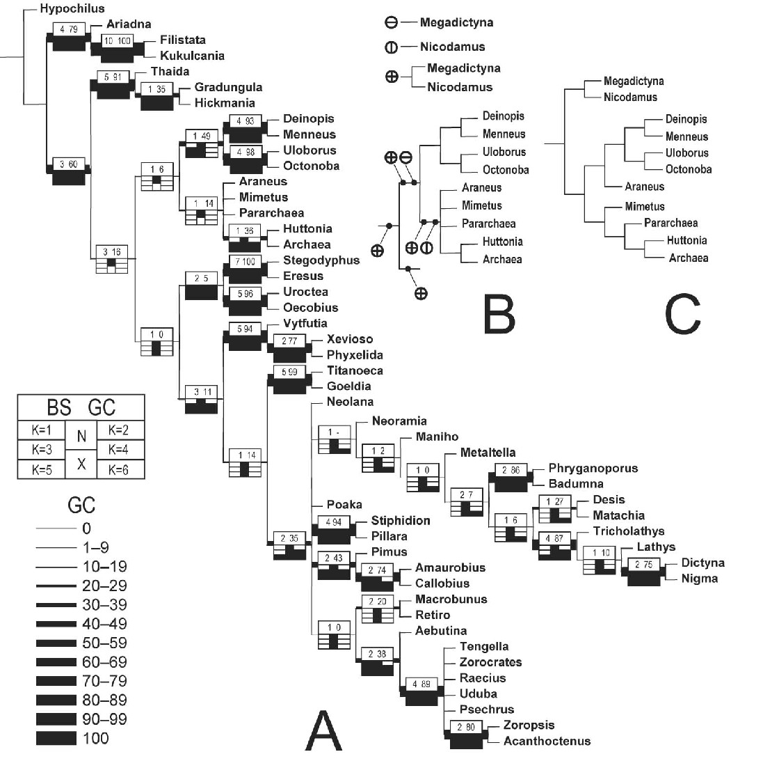
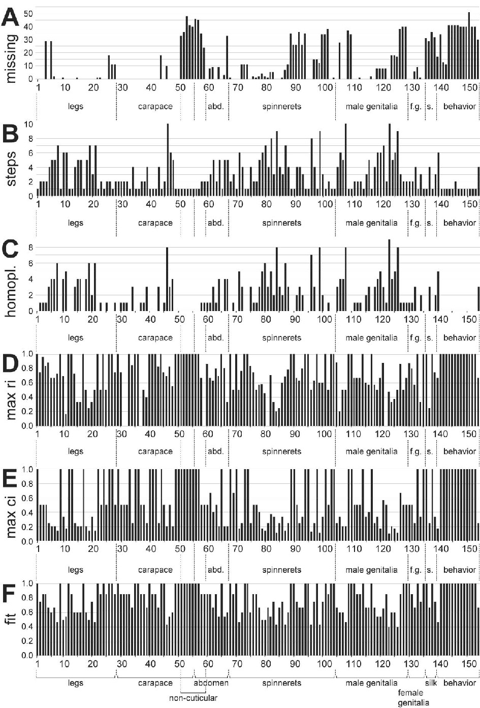